Transvaginal ultrasound assessment of corpus callosal length in the fetus: multicenter cross-sectional study.

IF 6.1 1区 医学 Q1 ACOUSTICS
D Paladini, S Parodi, H Xie, F Viñals, K Haratz, R Birnbaum, G Azumendi, L Pomar, E Montaguti, P Acharya, P Volpe, M Perez Cruz, K Katrin, R Chaoui, R Pooh
{"title":"Transvaginal ultrasound assessment of corpus callosal length in the fetus: multicenter cross-sectional study.","authors":"D Paladini, S Parodi, H Xie, F Viñals, K Haratz, R Birnbaum, G Azumendi, L Pomar, E Montaguti, P Acharya, P Volpe, M Perez Cruz, K Katrin, R Chaoui, R Pooh","doi":"10.1002/uog.29227","DOIUrl":null,"url":null,"abstract":"<p><strong>Objective: </strong>To produce reference ranges and Z-scores for corpus callosal (CC) length in the fetus, based on transvaginal three-dimensional (3D) ultrasound imaging.</p><p><strong>Methods: </strong>This was a cross-sectional multicenter retrospective study based on 3D volume dataset acquisitions of the fetal CC between the 15<sup>th</sup> and 37<sup>th</sup> weeks of gestation. Only volume datasets acquired transvaginally through the anterior fontanelle were selected. After plane alignment on multiplanar imaging, the length of the CC was measured edge-to-edge on magnified images. Intra- and interobserver variability were assessed and the related intraclass correlation coefficients (ICC) calculated. Biometric charts to assess the reference values for fetal CC were obtained using the method proposed by Altman in 1993.</p><p><strong>Results: </strong>The 13 participating centers provided valid data for 2131 patients. Excellent agreement was observed for both intra- and interobserver analysis, with an ICC range of 0.98-1.00. A quadratic model was used for construction of the reference charts, modified with the insertion of cubic spline coefficients with a single knot at 18 gestational weeks, to recover an apparent lack of fit at lower gestational ages. Centile reference values and the corresponding Z-scores were produced for CC length between 15 and 37 gestational weeks.</p><p><strong>Conclusions: </strong>This multicenter study presents growth charts for the fetal CC, addressing the critical methodological weaknesses of several previous studies. An even distribution of cases across all gestational weeks, robust statistical methodology and a standardized, high-resolution transvaginal neurosonographic technique represent key factors supporting the reliability of the biometric curves presented here. © 2025 The Author(s). Ultrasound in Obstetrics & Gynecology published by John Wiley & Sons Ltd on behalf of International Society of Ultrasound in Obstetrics and Gynecology.</p>","PeriodicalId":23454,"journal":{"name":"Ultrasound in Obstetrics & Gynecology","volume":" ","pages":""},"PeriodicalIF":6.1000,"publicationDate":"2025-05-05","publicationTypes":"Journal Article","fieldsOfStudy":null,"isOpenAccess":false,"openAccessPdf":"","citationCount":"0","resultStr":null,"platform":"Semanticscholar","paperid":null,"PeriodicalName":"Ultrasound in Obstetrics & Gynecology","FirstCategoryId":"3","ListUrlMain":"https://doi.org/10.1002/uog.29227","RegionNum":1,"RegionCategory":"医学","ArticlePicture":[],"TitleCN":null,"AbstractTextCN":null,"PMCID":null,"EPubDate":"","PubModel":"","JCR":"Q1","JCRName":"ACOUSTICS","Score":null,"Total":0}
引用次数: 0

Abstract

Objective: To produce reference ranges and Z-scores for corpus callosal (CC) length in the fetus, based on transvaginal three-dimensional (3D) ultrasound imaging.

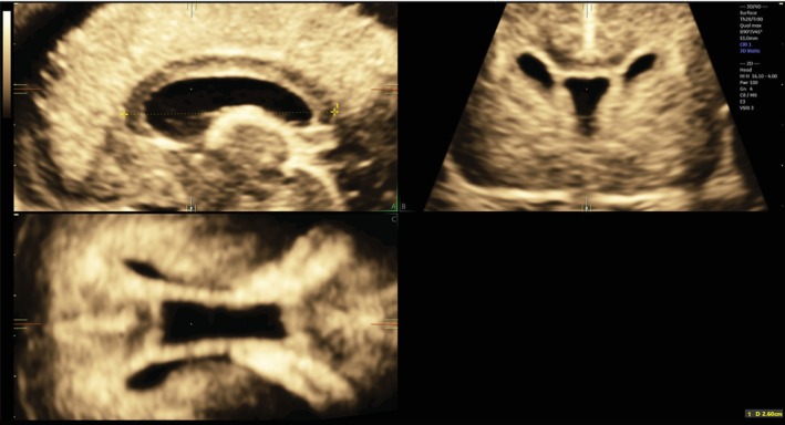
Methods: This was a cross-sectional multicenter retrospective study based on 3D volume dataset acquisitions of the fetal CC between the 15th and 37th weeks of gestation. Only volume datasets acquired transvaginally through the anterior fontanelle were selected. After plane alignment on multiplanar imaging, the length of the CC was measured edge-to-edge on magnified images. Intra- and interobserver variability were assessed and the related intraclass correlation coefficients (ICC) calculated. Biometric charts to assess the reference values for fetal CC were obtained using the method proposed by Altman in 1993.

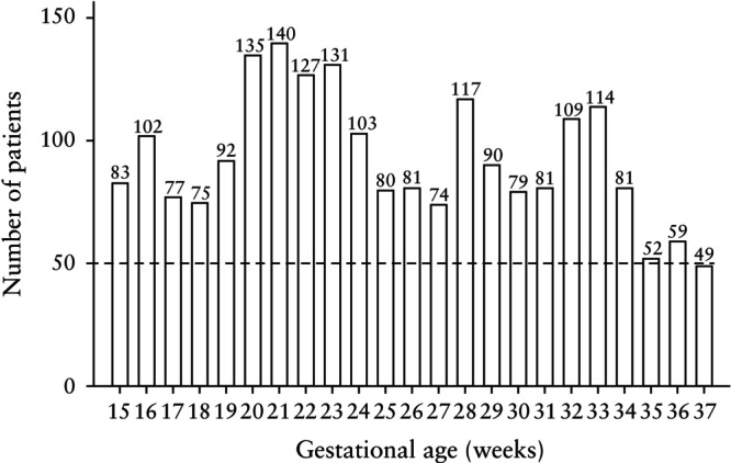
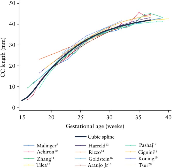
Results: The 13 participating centers provided valid data for 2131 patients. Excellent agreement was observed for both intra- and interobserver analysis, with an ICC range of 0.98-1.00. A quadratic model was used for construction of the reference charts, modified with the insertion of cubic spline coefficients with a single knot at 18 gestational weeks, to recover an apparent lack of fit at lower gestational ages. Centile reference values and the corresponding Z-scores were produced for CC length between 15 and 37 gestational weeks.

Conclusions: This multicenter study presents growth charts for the fetal CC, addressing the critical methodological weaknesses of several previous studies. An even distribution of cases across all gestational weeks, robust statistical methodology and a standardized, high-resolution transvaginal neurosonographic technique represent key factors supporting the reliability of the biometric curves presented here. © 2025 The Author(s). Ultrasound in Obstetrics & Gynecology published by John Wiley & Sons Ltd on behalf of International Society of Ultrasound in Obstetrics and Gynecology.

经阴道超声评估胎儿胼胝体长度:多中心横断面研究。
目的:基于经阴道三维超声成像,建立胎儿胼胝体(CC)长度的参考范围和z评分。方法:这是一项基于妊娠第15至37周胎儿CC三维体积数据集获取的横断面多中心回顾性研究。仅选择经阴道经前囟门获得的体积数据集。在多平面成像上平面对准后,在放大图像上逐边测量CC的长度。评估了观察者内部和观察者之间的变异,并计算了相关的类内相关系数(ICC)。采用Altman(1993)提出的方法获得生物特征图,以评估胎儿CC的参考值。结果:13个参与中心为2131例患者提供有效资料。在观察者内部和观察者之间的分析中观察到非常好的一致性,ICC范围为0.98-1.00。二次模型用于构建参考图表,并在18孕周时插入三次样条系数与单个结进行修改,以恢复低胎龄时明显缺乏拟合。在15至37孕周期间,计算CC长度的百分位数参考值和相应的z分数。结论:这项多中心研究提供了胎儿CC的生长图表,解决了以前几项研究的关键方法学弱点。所有妊娠周的病例均匀分布,稳健的统计方法和标准化的高分辨率经阴道神经超声技术是支持这里提出的生物特征曲线可靠性的关键因素。©2025作者。妇产科学超声由John Wiley & Sons Ltd代表国际妇产科学超声学会出版。
本文章由计算机程序翻译,如有差异,请以英文原文为准。
求助全文
约1分钟内获得全文 求助全文
来源期刊
CiteScore
12.30
自引率
14.10%
发文量
891
审稿时长
1 months
期刊介绍: Ultrasound in Obstetrics & Gynecology (UOG) is the official journal of the International Society of Ultrasound in Obstetrics and Gynecology (ISUOG) and is considered the foremost international peer-reviewed journal in the field. It publishes cutting-edge research that is highly relevant to clinical practice, which includes guidelines, expert commentaries, consensus statements, original articles, and systematic reviews. UOG is widely recognized and included in prominent abstract and indexing databases such as Index Medicus and Current Contents.
×
引用
GB/T 7714-2015
复制
MLA
复制
APA
复制
导出至
BibTeX EndNote RefMan NoteFirst NoteExpress
×
提示
您的信息不完整,为了账户安全,请先补充。
现在去补充
×
提示
您因"违规操作"
具体请查看互助需知
我知道了
×
提示
确定
请完成安全验证×
copy
已复制链接
快去分享给好友吧!
我知道了
右上角分享
点击右上角分享
0
联系我们:info@booksci.cn Book学术提供免费学术资源搜索服务,方便国内外学者检索中英文文献。致力于提供最便捷和优质的服务体验。 Copyright © 2023 布克学术 All rights reserved.
京ICP备2023020795号-1
ghs 京公网安备 11010802042870号
Book学术文献互助
Book学术文献互助群
群 号:481959085
Book学术官方微信