AGN1 local osteo-enhancement procedure increases proximal femur volumetric bone mineral density of women with post-menopausal osteoporosis as assessed by quantitative computed tomography analysis.
Michelle Chin, Ronald Hill, Bryan Huber, James Howe, Klaus Engelke
{"title":"AGN1 local osteo-enhancement procedure increases proximal femur volumetric bone mineral density of women with post-menopausal osteoporosis as assessed by quantitative computed tomography analysis.","authors":"Michelle Chin, Ronald Hill, Bryan Huber, James Howe, Klaus Engelke","doi":"10.1093/jbmrpl/ziaf036","DOIUrl":null,"url":null,"abstract":"<p><p>In this study, QCT was used to analyze the AGN1 Local Osteo-Enhancement Procedure (LOEP) as a treatment to form bone in the proximal femurs of patients with osteoporosis. Using this minimally invasive procedure, a resorbable triphasic AGN1 implant material was injected into the left femurs of 12 women with post-menopausal osteoporosis. Computed tomography scans were taken before treatment (baseline) and at 12 wk, 24 wk, and 5-7 yr after treatment. Quantitative computed tomography was used to investigate the resorption of AGN1 within the treated proximal femurs and to analyze the treatment's impact on integral, trabecular, and cortical bone. The untreated right femurs were used as controls. Data illustrated an increase in trabecular volumetric BMD (trab vBMD) of treated hips at all timepoints (baseline: 22 ± 21 mg/cm<sup>3</sup> vs 217 ± 56 mg/cm<sup>3</sup>, 161 ± 18 mg/cm<sup>3</sup>, and 121 ± 37 mg/cm<sup>3</sup> at 12-wk, 24-wk, and 5- to 7-yr timepoints, respectively), and an increase in integral vBMD of 65% at the 12-wk timepoint and 34% at the 5- to 7-yr timepoint. The increase in trab vBMD was observed in the location where the AGN1 implant material bolus was injected, and at the 5- to 7-yr timepoint, no significant BMD change was observed in the trabecular regions surrounding the original implantation zone (treated: 32 ± 16 mg/cm<sup>3</sup>, control: 31 ± 16 mg/cm<sup>3</sup>). This QCT study provides a more detailed understanding of the resorption and transformation of the AGN1 implant material into bone and supports, with some limitations, that the AGN1 LOEP treatment can locally increase trabecular bone density in weakened areas of the proximal femur where strength increase is most needed to reduce the risk of hip fragility fracture.</p>","PeriodicalId":14611,"journal":{"name":"JBMR Plus","volume":"9 5","pages":"ziaf036"},"PeriodicalIF":2.4000,"publicationDate":"2025-04-04","publicationTypes":"Journal Article","fieldsOfStudy":null,"isOpenAccess":false,"openAccessPdf":"https://www.ncbi.nlm.nih.gov/pmc/articles/PMC12035697/pdf/","citationCount":"0","resultStr":null,"platform":"Semanticscholar","paperid":null,"PeriodicalName":"JBMR Plus","FirstCategoryId":"1085","ListUrlMain":"https://doi.org/10.1093/jbmrpl/ziaf036","RegionNum":0,"RegionCategory":null,"ArticlePicture":[],"TitleCN":null,"AbstractTextCN":null,"PMCID":null,"EPubDate":"2025/5/1 0:00:00","PubModel":"eCollection","JCR":"Q2","JCRName":"ENDOCRINOLOGY & METABOLISM","Score":null,"Total":0}
引用次数: 0
Abstract
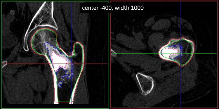
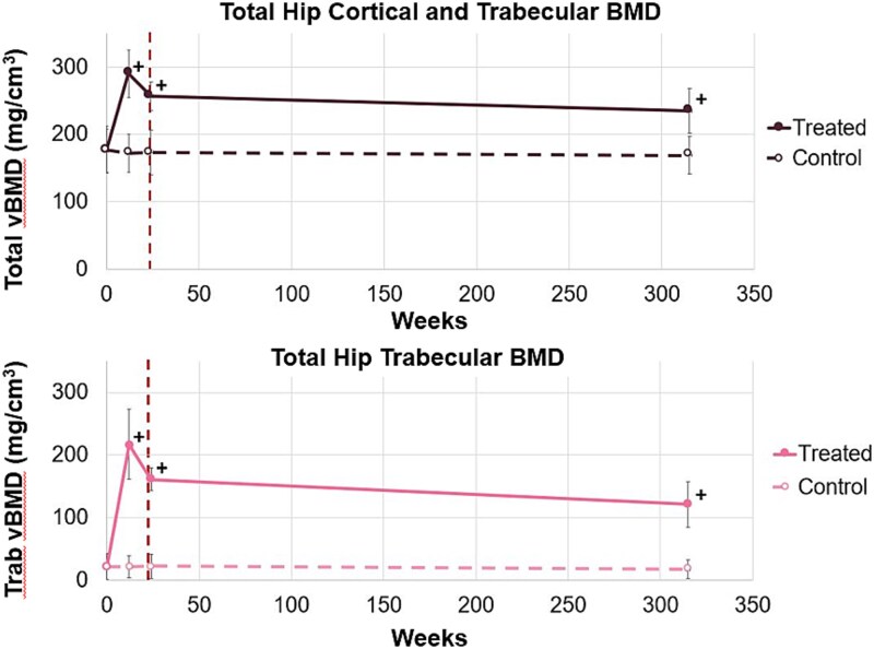
In this study, QCT was used to analyze the AGN1 Local Osteo-Enhancement Procedure (LOEP) as a treatment to form bone in the proximal femurs of patients with osteoporosis. Using this minimally invasive procedure, a resorbable triphasic AGN1 implant material was injected into the left femurs of 12 women with post-menopausal osteoporosis. Computed tomography scans were taken before treatment (baseline) and at 12 wk, 24 wk, and 5-7 yr after treatment. Quantitative computed tomography was used to investigate the resorption of AGN1 within the treated proximal femurs and to analyze the treatment's impact on integral, trabecular, and cortical bone. The untreated right femurs were used as controls. Data illustrated an increase in trabecular volumetric BMD (trab vBMD) of treated hips at all timepoints (baseline: 22 ± 21 mg/cm3 vs 217 ± 56 mg/cm3, 161 ± 18 mg/cm3, and 121 ± 37 mg/cm3 at 12-wk, 24-wk, and 5- to 7-yr timepoints, respectively), and an increase in integral vBMD of 65% at the 12-wk timepoint and 34% at the 5- to 7-yr timepoint. The increase in trab vBMD was observed in the location where the AGN1 implant material bolus was injected, and at the 5- to 7-yr timepoint, no significant BMD change was observed in the trabecular regions surrounding the original implantation zone (treated: 32 ± 16 mg/cm3, control: 31 ± 16 mg/cm3). This QCT study provides a more detailed understanding of the resorption and transformation of the AGN1 implant material into bone and supports, with some limitations, that the AGN1 LOEP treatment can locally increase trabecular bone density in weakened areas of the proximal femur where strength increase is most needed to reduce the risk of hip fragility fracture.


 求助内容:
求助内容: 应助结果提醒方式:


