Hanan Amer, Hanan Helmy, Enji El-Sawy, Maha S Ayoub, Nesma Mounir
{"title":"Parenchymal neuro-sonological characteristics in epileptic patients and their correlation with cognitive dysfunction.","authors":"Hanan Amer, Hanan Helmy, Enji El-Sawy, Maha S Ayoub, Nesma Mounir","doi":"10.1186/s42494-025-00212-8","DOIUrl":null,"url":null,"abstract":"<p><strong>Background: </strong>Idiopathic generalized epilepsies (IGEs) are the most common syndromes within the \"genetic generalized epilepsies\" (GGEs). Patients with IGE often exhibit cognitive comorbidities. The primary objective of this study is to investigate the correlation between brain parenchymal sonography characteristics and cognitive impairment in IGE.</p><p><strong>Methods: </strong>This study enrolled 26 patients with IGE and 26 age- and sex-matched controls. All participants underwent comprehensive evaluations including clinical examination, electroencephalography, magnetic resonance imaging epilepsy protocol, transcranial sonography (TCS) for third and lateral ventricular diameter measurements, and cognitive assessment using the Addenbrooke's Cognitive Examination-III (ACE III).</p><p><strong>Results: </strong>This study found significantly lower scores in attention, memory, fluency, and total score of ACE-III in IGE patients compared to the control group (P-value = 0.011, 0.033, 0.007, and 0.001, respectively). However, no significant differences were observed between IGE patients and the control group in language and visuospatial score (P = 0.479 and 0.108, respectively). The average diameters of the third ventricle and lateral ventricle anterior horns were significantly larger in patients than in the control group (P-value 0.004, 0.009, and 0.012, respectively).</p><p><strong>Conclusions: </strong>IGE patients exhibit significant cognitive impairment and notable dilatation of the third ventricle and lateral ventricles horns, which may serve as markers of brain atrophy.</p>","PeriodicalId":33628,"journal":{"name":"Acta Epileptologica","volume":"7 1","pages":"26"},"PeriodicalIF":1.2000,"publicationDate":"2025-04-21","publicationTypes":"Journal Article","fieldsOfStudy":null,"isOpenAccess":false,"openAccessPdf":"https://www.ncbi.nlm.nih.gov/pmc/articles/PMC12010533/pdf/","citationCount":"0","resultStr":null,"platform":"Semanticscholar","paperid":null,"PeriodicalName":"Acta Epileptologica","FirstCategoryId":"1085","ListUrlMain":"https://doi.org/10.1186/s42494-025-00212-8","RegionNum":0,"RegionCategory":null,"ArticlePicture":[],"TitleCN":null,"AbstractTextCN":null,"PMCID":null,"EPubDate":"","PubModel":"","JCR":"Q4","JCRName":"CLINICAL NEUROLOGY","Score":null,"Total":0}
引用次数: 0
Abstract
Background: Idiopathic generalized epilepsies (IGEs) are the most common syndromes within the "genetic generalized epilepsies" (GGEs). Patients with IGE often exhibit cognitive comorbidities. The primary objective of this study is to investigate the correlation between brain parenchymal sonography characteristics and cognitive impairment in IGE.
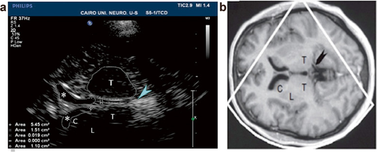
Methods: This study enrolled 26 patients with IGE and 26 age- and sex-matched controls. All participants underwent comprehensive evaluations including clinical examination, electroencephalography, magnetic resonance imaging epilepsy protocol, transcranial sonography (TCS) for third and lateral ventricular diameter measurements, and cognitive assessment using the Addenbrooke's Cognitive Examination-III (ACE III).
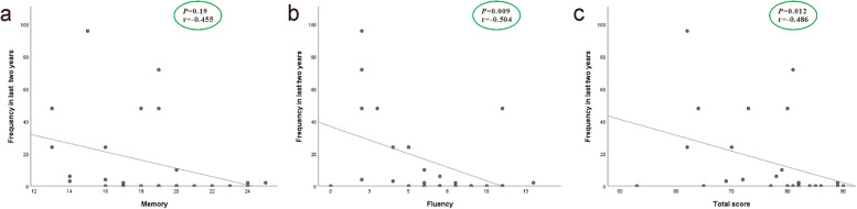
Results: This study found significantly lower scores in attention, memory, fluency, and total score of ACE-III in IGE patients compared to the control group (P-value = 0.011, 0.033, 0.007, and 0.001, respectively). However, no significant differences were observed between IGE patients and the control group in language and visuospatial score (P = 0.479 and 0.108, respectively). The average diameters of the third ventricle and lateral ventricle anterior horns were significantly larger in patients than in the control group (P-value 0.004, 0.009, and 0.012, respectively).
Conclusions: IGE patients exhibit significant cognitive impairment and notable dilatation of the third ventricle and lateral ventricles horns, which may serve as markers of brain atrophy.
 求助内容:
求助内容: 应助结果提醒方式:


