Yashar Ahmadyar, Alireza Kamali-Asl, Rezvan Samimi, Hossein Arabi, Habib Zaidi
{"title":"Automated pulmonary nodule classification from low-dose CT images using ERBNet: an ensemble learning approach.","authors":"Yashar Ahmadyar, Alireza Kamali-Asl, Rezvan Samimi, Hossein Arabi, Habib Zaidi","doi":"10.1007/s11517-025-03358-2","DOIUrl":null,"url":null,"abstract":"<p><p>The aim of this study was to develop a deep learning method for analyzing CT images with varying doses and qualities, aiming to categorize lung lesions into nodules and non-nodules. This study utilized the lung nodule analysis 2016 challenge dataset. Different low-dose CT (LDCT) images, including 10%, 20%, 40%, and 60% levels, were generated from the full-dose CT (FDCT) images. Five different 3D convolutional networks were developed to classify lung nodules from LDCT and reference FDCT images. The models were evaluated using 400 nodule and 400 non-nodule samples. An ensemble model was also developed to achieve a generalizable model across different dose levels. The model achieved an accuracy of 97.0% for nodule classification on FDCT images. However, the model exhibited relatively poor performance (60% accuracy) on LDCT images, indicating that dedicated models should be developed for each low-dose level. Dedicated models for handling LDCT led to dramatic increases in the accuracy of nodule classification. The dedicated low-dose models achieved a nodule classification accuracy of 90.0%, 91.1%, 92.7%, and 93.8% for 10%, 20%, 40%, and 60% of FDCT images, respectively. The accuracy of the deep learning models decreased gradually by almost 7% as LDCT images proceeded from 100 to 10%. However, the ensemble model led to an accuracy of 95.0% when tested on a combination of various dose levels. We presented an ensemble 3D CNN classifier for lesion classification, utilizing both LDCT and FDCT images. This model is able to analyze a combination of CT images with different dose levels and image qualities.</p>","PeriodicalId":49840,"journal":{"name":"Medical & Biological Engineering & Computing","volume":" ","pages":"2767-2779"},"PeriodicalIF":2.6000,"publicationDate":"2025-09-01","publicationTypes":"Journal Article","fieldsOfStudy":null,"isOpenAccess":false,"openAccessPdf":"https://www.ncbi.nlm.nih.gov/pmc/articles/PMC12402046/pdf/","citationCount":"0","resultStr":null,"platform":"Semanticscholar","paperid":null,"PeriodicalName":"Medical & Biological Engineering & Computing","FirstCategoryId":"5","ListUrlMain":"https://doi.org/10.1007/s11517-025-03358-2","RegionNum":4,"RegionCategory":"医学","ArticlePicture":[],"TitleCN":null,"AbstractTextCN":null,"PMCID":null,"EPubDate":"2025/4/15 0:00:00","PubModel":"Epub","JCR":"Q2","JCRName":"COMPUTER SCIENCE, INTERDISCIPLINARY APPLICATIONS","Score":null,"Total":0}
引用次数: 0
Abstract
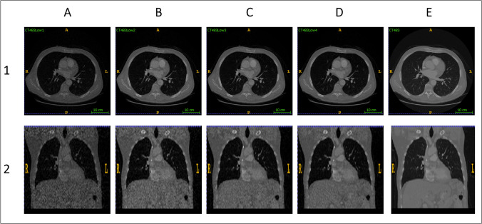
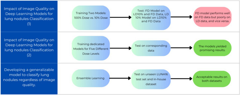
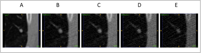
The aim of this study was to develop a deep learning method for analyzing CT images with varying doses and qualities, aiming to categorize lung lesions into nodules and non-nodules. This study utilized the lung nodule analysis 2016 challenge dataset. Different low-dose CT (LDCT) images, including 10%, 20%, 40%, and 60% levels, were generated from the full-dose CT (FDCT) images. Five different 3D convolutional networks were developed to classify lung nodules from LDCT and reference FDCT images. The models were evaluated using 400 nodule and 400 non-nodule samples. An ensemble model was also developed to achieve a generalizable model across different dose levels. The model achieved an accuracy of 97.0% for nodule classification on FDCT images. However, the model exhibited relatively poor performance (60% accuracy) on LDCT images, indicating that dedicated models should be developed for each low-dose level. Dedicated models for handling LDCT led to dramatic increases in the accuracy of nodule classification. The dedicated low-dose models achieved a nodule classification accuracy of 90.0%, 91.1%, 92.7%, and 93.8% for 10%, 20%, 40%, and 60% of FDCT images, respectively. The accuracy of the deep learning models decreased gradually by almost 7% as LDCT images proceeded from 100 to 10%. However, the ensemble model led to an accuracy of 95.0% when tested on a combination of various dose levels. We presented an ensemble 3D CNN classifier for lesion classification, utilizing both LDCT and FDCT images. This model is able to analyze a combination of CT images with different dose levels and image qualities.
期刊介绍:
Founded in 1963, Medical & Biological Engineering & Computing (MBEC) continues to serve the biomedical engineering community, covering the entire spectrum of biomedical and clinical engineering. The journal presents exciting and vital experimental and theoretical developments in biomedical science and technology, and reports on advances in computer-based methodologies in these multidisciplinary subjects. The journal also incorporates new and evolving technologies including cellular engineering and molecular imaging.
MBEC publishes original research articles as well as reviews and technical notes. Its Rapid Communications category focuses on material of immediate value to the readership, while the Controversies section provides a forum to exchange views on selected issues, stimulating a vigorous and informed debate in this exciting and high profile field.
MBEC is an official journal of the International Federation of Medical and Biological Engineering (IFMBE).

 求助内容:
求助内容: 应助结果提醒方式:


