Noor Fazaldad, Srinivasa Rao Sirasanagandla, Anwar Al-Shuaili, Sreenivasulu Reddy Mogali, Ramya Chandrasekaran, Humoud Al Dhuhli, Eiman Al-Ajmi
{"title":"Anatomical Variations and Morphometry of Carotid Sinus: A Computed Tomography Study.","authors":"Noor Fazaldad, Srinivasa Rao Sirasanagandla, Anwar Al-Shuaili, Sreenivasulu Reddy Mogali, Ramya Chandrasekaran, Humoud Al Dhuhli, Eiman Al-Ajmi","doi":"10.3390/tomography11040045","DOIUrl":null,"url":null,"abstract":"<p><strong>Background: </strong>The radiological evaluation of the carotid sinus (CS) anatomy and its morphometry is essentially important for various surgical procedures involving the carotid bifurcation and the CS itself. Despite its tremendous clinical significance, studies dealing with the CS anatomy are seldom reported. Hence, the present study aimed to evaluate the frequencies of the CS positional variants and their morphometry and correlate them with age and body mass index (BMI).</p><p><strong>Methods: </strong>In this retrospective cross-sectional study, a total of 754 disease-free carotid arteries were examined using computed tomography angiography scans to determine the CS positional variations (such as types I to III) and its morphometry, including the CS diameter and length. Additionally, the association between these parameters and factors such as sex, age, and body mass index were explored using appropriate statistical tests. The inter-rater agreement of the collected dataset was evaluated using Cohen's Kappa.</p><p><strong>Results: </strong>The CS type I was observed in 87.67% of the cases, and type II and type III were observed at lower frequencies with 9.02% and 3.32%, respectively. There were statistically significant (<i>p</i> < 0.001) differences observed in the mean diameter and length of the sinus between the sex and the type I CS variations. However, there was no significant and strong correlation between the age and BMI factors with sinus length and sinus diameter. The kappa values for inter-rater agreement ranged from 0.77 to 0.99 for all parameters.</p><p><strong>Conclusions: </strong>In type I, the CS length and carotid vessel's diameter is significantly different between the sexes. However, age and BMI do not affect the CS anatomy in radiologically disease-free carotid arteries. Knowledge of the CS variant anatomy is clinically significant as it influences the patients' surgical and physiological outcomes.</p>","PeriodicalId":51330,"journal":{"name":"Tomography","volume":"11 4","pages":""},"PeriodicalIF":2.2000,"publicationDate":"2025-04-07","publicationTypes":"Journal Article","fieldsOfStudy":null,"isOpenAccess":false,"openAccessPdf":"https://www.ncbi.nlm.nih.gov/pmc/articles/PMC12031040/pdf/","citationCount":"0","resultStr":null,"platform":"Semanticscholar","paperid":null,"PeriodicalName":"Tomography","FirstCategoryId":"3","ListUrlMain":"https://doi.org/10.3390/tomography11040045","RegionNum":4,"RegionCategory":"医学","ArticlePicture":[],"TitleCN":null,"AbstractTextCN":null,"PMCID":null,"EPubDate":"","PubModel":"","JCR":"Q2","JCRName":"RADIOLOGY, NUCLEAR MEDICINE & MEDICAL IMAGING","Score":null,"Total":0}
引用次数: 0
Abstract
Background: The radiological evaluation of the carotid sinus (CS) anatomy and its morphometry is essentially important for various surgical procedures involving the carotid bifurcation and the CS itself. Despite its tremendous clinical significance, studies dealing with the CS anatomy are seldom reported. Hence, the present study aimed to evaluate the frequencies of the CS positional variants and their morphometry and correlate them with age and body mass index (BMI).
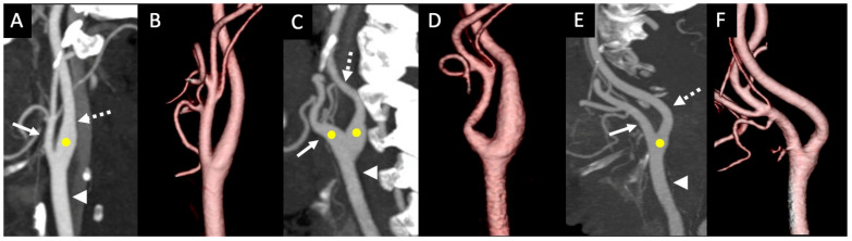
Methods: In this retrospective cross-sectional study, a total of 754 disease-free carotid arteries were examined using computed tomography angiography scans to determine the CS positional variations (such as types I to III) and its morphometry, including the CS diameter and length. Additionally, the association between these parameters and factors such as sex, age, and body mass index were explored using appropriate statistical tests. The inter-rater agreement of the collected dataset was evaluated using Cohen's Kappa.
Results: The CS type I was observed in 87.67% of the cases, and type II and type III were observed at lower frequencies with 9.02% and 3.32%, respectively. There were statistically significant (p < 0.001) differences observed in the mean diameter and length of the sinus between the sex and the type I CS variations. However, there was no significant and strong correlation between the age and BMI factors with sinus length and sinus diameter. The kappa values for inter-rater agreement ranged from 0.77 to 0.99 for all parameters.
Conclusions: In type I, the CS length and carotid vessel's diameter is significantly different between the sexes. However, age and BMI do not affect the CS anatomy in radiologically disease-free carotid arteries. Knowledge of the CS variant anatomy is clinically significant as it influences the patients' surgical and physiological outcomes.
TomographyMedicine-Radiology, Nuclear Medicine and Imaging
CiteScore
2.70
自引率
10.50%
发文量
222
期刊介绍:
TomographyTM publishes basic (technical and pre-clinical) and clinical scientific articles which involve the advancement of imaging technologies. Tomography encompasses studies that use single or multiple imaging modalities including for example CT, US, PET, SPECT, MR and hyperpolarization technologies, as well as optical modalities (i.e. bioluminescence, photoacoustic, endomicroscopy, fiber optic imaging and optical computed tomography) in basic sciences, engineering, preclinical and clinical medicine.
Tomography also welcomes studies involving exploration and refinement of contrast mechanisms and image-derived metrics within and across modalities toward the development of novel imaging probes for image-based feedback and intervention. The use of imaging in biology and medicine provides unparalleled opportunities to noninvasively interrogate tissues to obtain real-time dynamic and quantitative information required for diagnosis and response to interventions and to follow evolving pathological conditions. As multi-modal studies and the complexities of imaging technologies themselves are ever increasing to provide advanced information to scientists and clinicians.
Tomography provides a unique publication venue allowing investigators the opportunity to more precisely communicate integrated findings related to the diverse and heterogeneous features associated with underlying anatomical, physiological, functional, metabolic and molecular genetic activities of normal and diseased tissue. Thus Tomography publishes peer-reviewed articles which involve the broad use of imaging of any tissue and disease type including both preclinical and clinical investigations. In addition, hardware/software along with chemical and molecular probe advances are welcome as they are deemed to significantly contribute towards the long-term goal of improving the overall impact of imaging on scientific and clinical discovery.

 求助内容:
求助内容: 应助结果提醒方式:


