Dhivakaran Gengatharan, Walter Soon Yaw Wong, Lee Kai Lin, John Wen Cong Thng, Huang Yilun
{"title":"Electromagnetic Navigation in Biportal Endoscopic Lumbar Spine Surgery.","authors":"Dhivakaran Gengatharan, Walter Soon Yaw Wong, Lee Kai Lin, John Wen Cong Thng, Huang Yilun","doi":"10.22603/ssrr.2024-0257","DOIUrl":null,"url":null,"abstract":"<p><strong>Introduction: </strong>Endoscopic Spine Surgery (ESS) has begun to gain traction as an alternative to traditional microscopic spine surgery, particularly for lumbar decompression. However, one of the challenges associated with this approach is the steep learning curve. A recent advancement in this field aims to flatten the learning curve by incorporating navigation into ESS. This technology provides valuable information on the extent of decompression, confirms the working level, and reduces radiation exposure.</p><p><strong>Technical note: </strong>We aimed to describe our experience using electromagnetic navigation in biportal endoscopic spine surgery (BESS). The surgical technique is initiated by positioning the patient prone on a radiolucent table. The navigation field generator is positioned over the caudal end of the patient. The navigation system is set up with patient mappers at the desired working levels. The patient tracker is implanted. The final fluoroscopy images are captured in anteroposterior and lateral views. Subsequently, standard incisions are made, and endoscopic decompression is performed. When required, various instruments can be used to confirm the level, angulation, and extent of decompression.</p><p><strong>Conclusions: </strong>Our experience showed that this approach reduced the need for intraoperative imaging and provided an accurate alternative to repeated intraoperative imaging. However, it does involve a significantly long setup. Further trials of larger scale are required to determine its efficacy.</p>","PeriodicalId":22253,"journal":{"name":"Spine Surgery and Related Research","volume":"9 2","pages":"258-262"},"PeriodicalIF":1.2000,"publicationDate":"2024-12-20","publicationTypes":"Journal Article","fieldsOfStudy":null,"isOpenAccess":false,"openAccessPdf":"https://www.ncbi.nlm.nih.gov/pmc/articles/PMC11983126/pdf/","citationCount":"0","resultStr":null,"platform":"Semanticscholar","paperid":null,"PeriodicalName":"Spine Surgery and Related Research","FirstCategoryId":"1085","ListUrlMain":"https://doi.org/10.22603/ssrr.2024-0257","RegionNum":0,"RegionCategory":null,"ArticlePicture":[],"TitleCN":null,"AbstractTextCN":null,"PMCID":null,"EPubDate":"2025/3/27 0:00:00","PubModel":"eCollection","JCR":"Q3","JCRName":"SURGERY","Score":null,"Total":0}
引用次数: 0
Abstract
Introduction: Endoscopic Spine Surgery (ESS) has begun to gain traction as an alternative to traditional microscopic spine surgery, particularly for lumbar decompression. However, one of the challenges associated with this approach is the steep learning curve. A recent advancement in this field aims to flatten the learning curve by incorporating navigation into ESS. This technology provides valuable information on the extent of decompression, confirms the working level, and reduces radiation exposure.
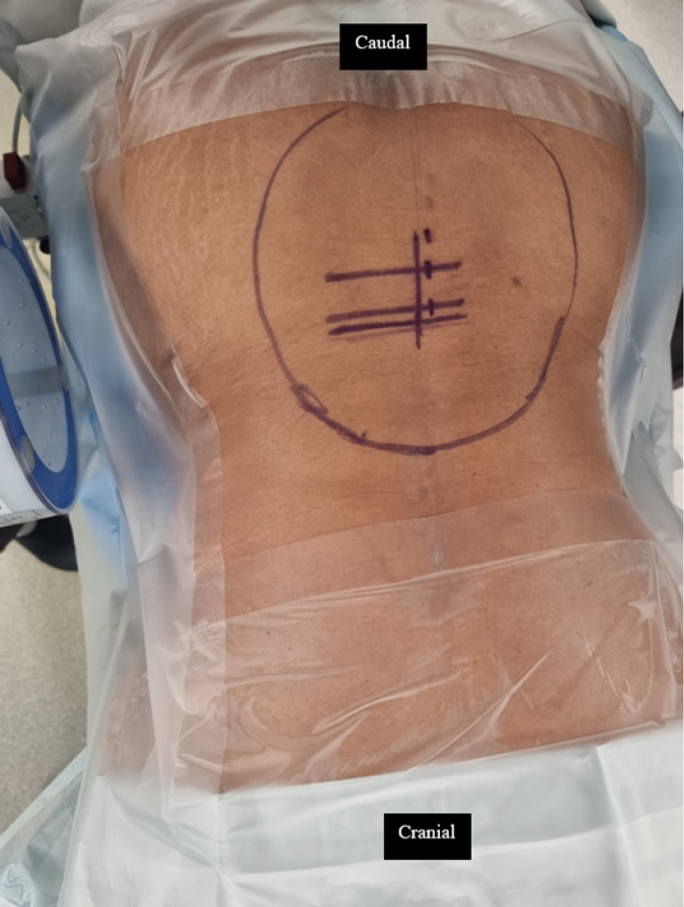
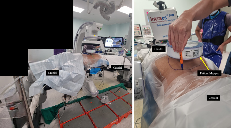
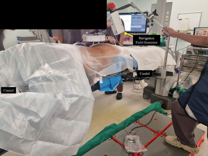
Technical note: We aimed to describe our experience using electromagnetic navigation in biportal endoscopic spine surgery (BESS). The surgical technique is initiated by positioning the patient prone on a radiolucent table. The navigation field generator is positioned over the caudal end of the patient. The navigation system is set up with patient mappers at the desired working levels. The patient tracker is implanted. The final fluoroscopy images are captured in anteroposterior and lateral views. Subsequently, standard incisions are made, and endoscopic decompression is performed. When required, various instruments can be used to confirm the level, angulation, and extent of decompression.
Conclusions: Our experience showed that this approach reduced the need for intraoperative imaging and provided an accurate alternative to repeated intraoperative imaging. However, it does involve a significantly long setup. Further trials of larger scale are required to determine its efficacy.

 求助内容:
求助内容: 应助结果提醒方式:


