Morphological and autofluorescence assessment of oocysts differentiate live from dead coccidian parasites
IF 3.2
2区 医学
Q1 PARASITOLOGY
引用次数: 0
Abstract
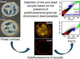
To assess and mitigate foodborne risk, regulatory agencies and produce growers require the means not merely to detect but moreover determine the viability of foodborne eukaryotic pathogens such as Cyclospora cayetanensis. Viability assessment would also aid those employing live attenuated vaccines against coccidiosis, a major problem in poultry production. Therefore, we sought to identify morphological changes differentiating viable from non-viable coccidian oocysts, employing Eimeria acervulina as a tractable model, enabling empirical validation by means of in vivo challenge infections in the natural chicken host. High resolution microscopic examinations identified granular structures that autofluoresce under UV exposure in dead oocysts, greatly intensifying overall autofluorescence in dead oocysts. We harnessed this intensification as a basis to sort live from dead oocysts using a Fluorescence-Activated Cell Sorting (FACS) cell sorter, validating their distinction by documenting infectivity in chickens using the former, and minimal shedding with the latter. Our rapid, sensitive, and robust assay holds promise for application to other species of coccidia, including those important to livestock and public health.
卵囊形态和自身荧光鉴定区分活的和死的球虫寄生虫。
为了评估和减轻食源性风险,监管机构和农产品种植者不仅需要检测,而且还需要确定食源性真核病原体(如卡耶坦环孢子虫)的生存能力。生存能力评估也将有助于那些采用球虫病减毒活疫苗的人,球虫病是家禽生产中的一个主要问题。因此,我们试图确定区分有活力和无活力球虫卵囊的形态学变化,采用艾美耳球虫作为可处理的模型,通过在天然鸡宿主体内攻击感染的方式进行经验验证。高分辨率显微镜检查发现,死卵囊在紫外线照射下会发出自身荧光的颗粒结构,大大增强了死卵囊的整体自身荧光。我们利用这种强化作为基础,使用荧光激活细胞分选(FACS)细胞分选器对活卵囊和死卵囊进行分类,通过记录前者在鸡中的感染性和后者的最小脱落来验证它们的区别。我们的快速、灵敏、可靠的检测方法有望应用于其他种类的球虫,包括那些对牲畜和公众健康重要的球虫。
本文章由计算机程序翻译,如有差异,请以英文原文为准。
求助全文
约1分钟内获得全文
求助全文
来源期刊
CiteScore
8.40
自引率
2.50%
发文量
76
审稿时长
23 days
期刊介绍:
International Journal for Parasitology offers authors the option to sponsor nonsubscriber access to their articles on Elsevier electronic publishing platforms. For more information please view our Sponsored Articles page. The International Journal for Parasitology publishes the results of original research in all aspects of basic and applied parasitology, including all the fields covered by its Specialist Editors, and ranging from parasites and host-parasite relationships of intrinsic biological interest to those of social and economic importance in human and veterinary medicine and agriculture.
 求助内容:
求助内容: 应助结果提醒方式:


