Ajay K Mali, Sivasubramanian Murugappan, Jayashree Rajesh Prasad, Syed A M Tofail, Nanasaheb D Thorat
{"title":"A deep learning pipeline for morphological and viability assessment of 3D cancer cell spheroids.","authors":"Ajay K Mali, Sivasubramanian Murugappan, Jayashree Rajesh Prasad, Syed A M Tofail, Nanasaheb D Thorat","doi":"10.1093/biomethods/bpaf030","DOIUrl":null,"url":null,"abstract":"<p><p>Three-dimensional (3D) spheroid models have advanced cancer research by better mimicking the tumour microenvironment compared to traditional <b>two-</b>dimensional cell cultures. However, challenges persist in high-throughput analysis of morphological characteristics and cell viability, as traditional methods like manual fluorescence analysis are labour-intensive and inconsistent. Existing AI-based approaches often address segmentation or classification in isolation, lacking an integrated workflow. We propose a scalable, two-stage deep learning pipeline to address these gaps: (i) a U-Net model for precise detection and segmentation of 3D spheroids from microscopic images, achieving 95% prediction accuracy, and (ii) a CNN Regression Hybrid method for estimating live/dead cell percentages and classifying spheroids, with an <math> <mrow> <msup><mrow><mi>R</mi></mrow> <mrow><mn>2</mn></mrow> </msup> </mrow> </math> value of 98%. This end-to-end pipeline automates cell viability quantification and generates key morphological parameters for spheroid growth kinetics. By integrating segmentation and analysis, our method addresses environmental variability and morphological characterization challenges, offering a robust tool for drug discovery, toxicity screening, and clinical research. This approach significantly improves efficiency and scalability of 3D spheroid evaluations, paving the way for advancements in cancer therapeutics.</p>","PeriodicalId":36528,"journal":{"name":"Biology Methods and Protocols","volume":"10 1","pages":"bpaf030"},"PeriodicalIF":1.3000,"publicationDate":"2025-04-11","publicationTypes":"Journal Article","fieldsOfStudy":null,"isOpenAccess":false,"openAccessPdf":"https://www.ncbi.nlm.nih.gov/pmc/articles/PMC12064216/pdf/","citationCount":"0","resultStr":null,"platform":"Semanticscholar","paperid":null,"PeriodicalName":"Biology Methods and Protocols","FirstCategoryId":"1085","ListUrlMain":"https://doi.org/10.1093/biomethods/bpaf030","RegionNum":0,"RegionCategory":null,"ArticlePicture":[],"TitleCN":null,"AbstractTextCN":null,"PMCID":null,"EPubDate":"2025/1/1 0:00:00","PubModel":"eCollection","JCR":"Q3","JCRName":"BIOCHEMICAL RESEARCH METHODS","Score":null,"Total":0}
引用次数: 0
Abstract
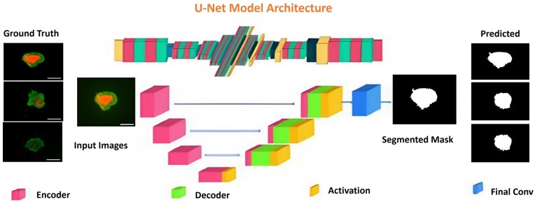
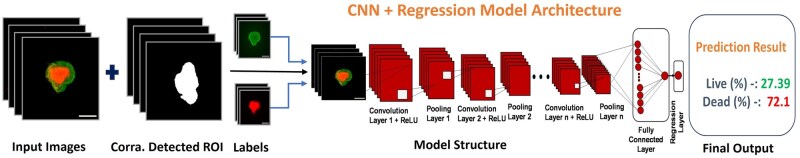
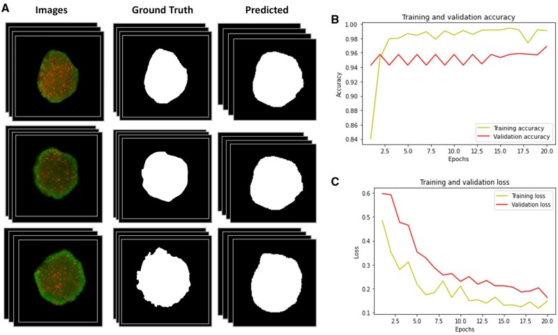
Three-dimensional (3D) spheroid models have advanced cancer research by better mimicking the tumour microenvironment compared to traditional two-dimensional cell cultures. However, challenges persist in high-throughput analysis of morphological characteristics and cell viability, as traditional methods like manual fluorescence analysis are labour-intensive and inconsistent. Existing AI-based approaches often address segmentation or classification in isolation, lacking an integrated workflow. We propose a scalable, two-stage deep learning pipeline to address these gaps: (i) a U-Net model for precise detection and segmentation of 3D spheroids from microscopic images, achieving 95% prediction accuracy, and (ii) a CNN Regression Hybrid method for estimating live/dead cell percentages and classifying spheroids, with an value of 98%. This end-to-end pipeline automates cell viability quantification and generates key morphological parameters for spheroid growth kinetics. By integrating segmentation and analysis, our method addresses environmental variability and morphological characterization challenges, offering a robust tool for drug discovery, toxicity screening, and clinical research. This approach significantly improves efficiency and scalability of 3D spheroid evaluations, paving the way for advancements in cancer therapeutics.

 求助内容:
求助内容: 应助结果提醒方式:


