Tamás F Polgár, Krisztina Spisák, Zalán Kádár, Nora Alodah, Gabor J Szebeni, Kata Klein, Roland Patai, László Siklós, Bernát Nógrádi
{"title":"Photobleaching alters the morphometric analysis of fluorescently labeled neurons and microglial cells.","authors":"Tamás F Polgár, Krisztina Spisák, Zalán Kádár, Nora Alodah, Gabor J Szebeni, Kata Klein, Roland Patai, László Siklós, Bernát Nógrádi","doi":"10.3389/pore.2025.1612087","DOIUrl":null,"url":null,"abstract":"<p><p>Photobleaching of immunofluorescence signal is a well-known phenomenon, however, its impact on derived parameters characterizing number and shape of different cell types in tissue sections is less understood. Our aim was to determine whether the duration of illumination and the type of fluorophore (Alexa Fluor 546 (A546), and Alexa Fluor 488 Plus (A488)) can influence the acquired morphometric parameters of cells in the nervous system. Immunofluorescent staining of microglia and neurons was performed on mouse spinal cord sections. Mean color intensity in a field of view, number of detectable neuronal cell profiles, partial coverage of microglial profiles, and fractal geometrical parameters were determined. All measurements were made using epifluorescence microscopy with identical acquisition parameters. Most of the measured parameters suffered significant alternation after 30-60 s of illumination. The data-altering effect of photobleaching was most prominent in the case of mean fluorescent intensity. Thus, while immunofluorescent staining is useful for co-localizing different groups of cells, cell-specific quantitative morphological measurements require photostable staining. Possibility of the combination of these methods on the same section in order to achieve multi-channel localization without photobleaching is exemplified.</p>","PeriodicalId":19981,"journal":{"name":"Pathology & Oncology Research","volume":"31 ","pages":"1612087"},"PeriodicalIF":2.3000,"publicationDate":"2025-04-04","publicationTypes":"Journal Article","fieldsOfStudy":null,"isOpenAccess":false,"openAccessPdf":"https://www.ncbi.nlm.nih.gov/pmc/articles/PMC12006003/pdf/","citationCount":"0","resultStr":null,"platform":"Semanticscholar","paperid":null,"PeriodicalName":"Pathology & Oncology Research","FirstCategoryId":"3","ListUrlMain":"https://doi.org/10.3389/pore.2025.1612087","RegionNum":4,"RegionCategory":"医学","ArticlePicture":[],"TitleCN":null,"AbstractTextCN":null,"PMCID":null,"EPubDate":"2025/1/1 0:00:00","PubModel":"eCollection","JCR":"Q3","JCRName":"ONCOLOGY","Score":null,"Total":0}
引用次数: 0
Abstract
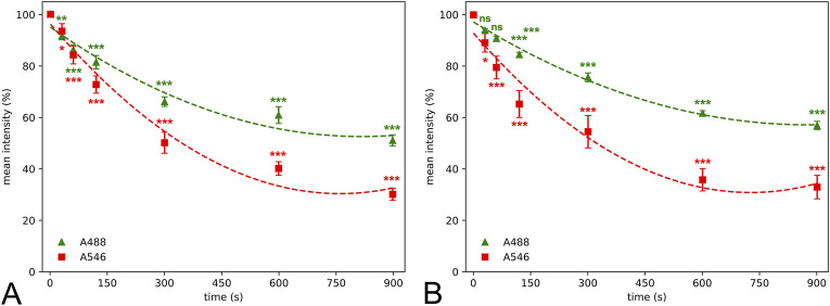
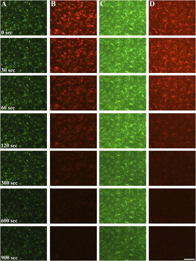
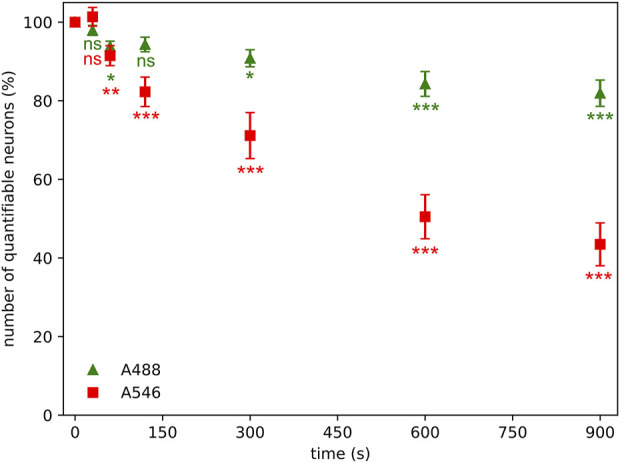
Photobleaching of immunofluorescence signal is a well-known phenomenon, however, its impact on derived parameters characterizing number and shape of different cell types in tissue sections is less understood. Our aim was to determine whether the duration of illumination and the type of fluorophore (Alexa Fluor 546 (A546), and Alexa Fluor 488 Plus (A488)) can influence the acquired morphometric parameters of cells in the nervous system. Immunofluorescent staining of microglia and neurons was performed on mouse spinal cord sections. Mean color intensity in a field of view, number of detectable neuronal cell profiles, partial coverage of microglial profiles, and fractal geometrical parameters were determined. All measurements were made using epifluorescence microscopy with identical acquisition parameters. Most of the measured parameters suffered significant alternation after 30-60 s of illumination. The data-altering effect of photobleaching was most prominent in the case of mean fluorescent intensity. Thus, while immunofluorescent staining is useful for co-localizing different groups of cells, cell-specific quantitative morphological measurements require photostable staining. Possibility of the combination of these methods on the same section in order to achieve multi-channel localization without photobleaching is exemplified.
期刊介绍:
Pathology & Oncology Research (POR) is an interdisciplinary Journal at the interface of pathology and oncology including the preclinical and translational research, diagnostics and therapy. Furthermore, POR is an international forum for the rapid communication of reviews, original research, critical and topical reports with excellence and novelty. Published quarterly, POR is dedicated to keeping scientists informed of developments on the selected biomedical fields bridging the gap between basic research and clinical medicine. It is a special aim for POR to promote pathological and oncological publishing activity of colleagues in the Central and East European region. The journal will be of interest to pathologists, and a broad range of experimental and clinical oncologists, and related experts. POR is supported by an acknowledged international advisory board and the Arányi Fundation for modern pathology.
 求助内容:
求助内容: 应助结果提醒方式:


