{"title":"Traumatic isolated spinous process fractures.","authors":"Muhittin Emre Altunrende, Elif Evrim Ekin","doi":"10.14744/tjtes.2025.20830","DOIUrl":null,"url":null,"abstract":"<p><strong>Background: </strong>Isolated traumatic spinous process fractures account for a small proportion of diagnosed spinal fractures. Among spinal fractures, the cervical vertebra is the most common site of involvement, often referred to as a Clay-Shoveler's fracture. This study included patients with isolated spinous process fractures through radiologic examinations over the past five years. The study aimed to investigate the cause of trauma, the presence of spinal ligament injury, and the relationship between these fractures with age and sex.</p><p><strong>Methods: </strong>Magnetic resonance imaging (MRI) and computed tomography (CT) scans of the spine, performed for any reason over the past five years at the hospital where the study was conducted, were retrospectively reviewed and re-evaluated. Patients whose examinations were unrelated to trauma, those with imaging artifacts that interfered with evaluation, and those who had undergone spinal vertebral surgery were excluded. The etiology of fractures was classified using the International Classification of Diseases (ICD) diagnoses. Data on age, sex, cause of trauma, and spinal ligament injury were recorded.</p><p><strong>Results: </strong>A total of 44 patients (36 men and eight women) were included in the study, with a mean age of 43.1 years. Among them, 25 patients were admitted due to traffic accidents and 18 due to falls. Spinous process fractures were observed in 18, 17, and three patients in the cervical, thoracic, and lumbar regions, respectively. Multiple spinous process fractures were found in 15 patients, while six patients had fractures in both the cervical and thoracic regions (Clay-Shoveler's fracture). In 12 patients, spinal MRI with Short-TI Inversion Recovery (STIR) sequences was performed in addition to CT imaging. All patients with Clay-Shoveler's fracture fractures underwent both CT and MRI examinations. In all cases where MRI was performed, interspinous ligament damage was detected. However, no intracanal involvement or comorbid pathology was observed.</p><p><strong>Conclusion: </strong>Notably, multiple fractures may occur, particularly at the cervicothoracic junction. Although spinous process fractures associated with trauma are rarely isolated, they are usually managed with medical treatment. Therefore, the vertebrae below the initially detected fracture site should also be evaluated. Additionally, imaging studies such as MRI with STIR sequences should be performed to assess ligament damage and the neural canal, in addition to tests for evaluating bony structures.</p>","PeriodicalId":94263,"journal":{"name":"Ulusal travma ve acil cerrahi dergisi = Turkish journal of trauma & emergency surgery : TJTES","volume":"31 4","pages":"394-398"},"PeriodicalIF":1.0000,"publicationDate":"2025-04-01","publicationTypes":"Journal Article","fieldsOfStudy":null,"isOpenAccess":false,"openAccessPdf":"https://www.ncbi.nlm.nih.gov/pmc/articles/PMC12000981/pdf/","citationCount":"0","resultStr":null,"platform":"Semanticscholar","paperid":null,"PeriodicalName":"Ulusal travma ve acil cerrahi dergisi = Turkish journal of trauma & emergency surgery : TJTES","FirstCategoryId":"1085","ListUrlMain":"https://doi.org/10.14744/tjtes.2025.20830","RegionNum":0,"RegionCategory":null,"ArticlePicture":[],"TitleCN":null,"AbstractTextCN":null,"PMCID":null,"EPubDate":"","PubModel":"","JCR":"","JCRName":"","Score":null,"Total":0}
引用次数: 0
Abstract
Background: Isolated traumatic spinous process fractures account for a small proportion of diagnosed spinal fractures. Among spinal fractures, the cervical vertebra is the most common site of involvement, often referred to as a Clay-Shoveler's fracture. This study included patients with isolated spinous process fractures through radiologic examinations over the past five years. The study aimed to investigate the cause of trauma, the presence of spinal ligament injury, and the relationship between these fractures with age and sex.
Methods: Magnetic resonance imaging (MRI) and computed tomography (CT) scans of the spine, performed for any reason over the past five years at the hospital where the study was conducted, were retrospectively reviewed and re-evaluated. Patients whose examinations were unrelated to trauma, those with imaging artifacts that interfered with evaluation, and those who had undergone spinal vertebral surgery were excluded. The etiology of fractures was classified using the International Classification of Diseases (ICD) diagnoses. Data on age, sex, cause of trauma, and spinal ligament injury were recorded.
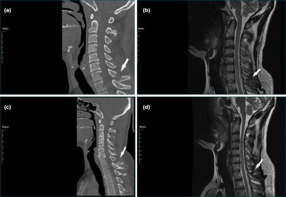
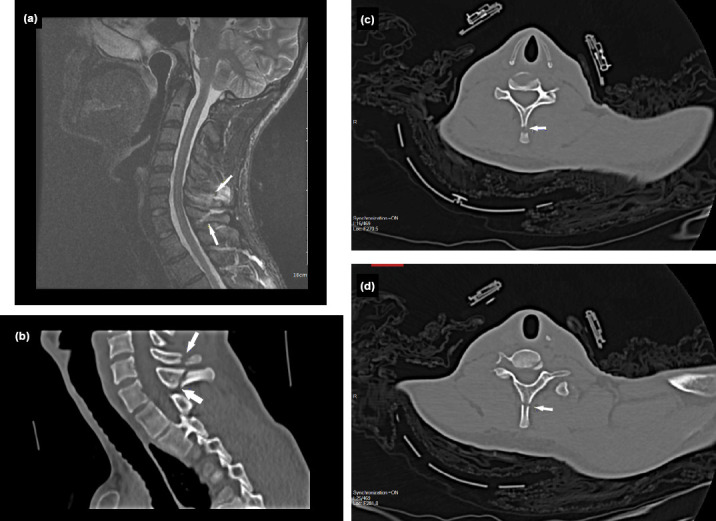
Results: A total of 44 patients (36 men and eight women) were included in the study, with a mean age of 43.1 years. Among them, 25 patients were admitted due to traffic accidents and 18 due to falls. Spinous process fractures were observed in 18, 17, and three patients in the cervical, thoracic, and lumbar regions, respectively. Multiple spinous process fractures were found in 15 patients, while six patients had fractures in both the cervical and thoracic regions (Clay-Shoveler's fracture). In 12 patients, spinal MRI with Short-TI Inversion Recovery (STIR) sequences was performed in addition to CT imaging. All patients with Clay-Shoveler's fracture fractures underwent both CT and MRI examinations. In all cases where MRI was performed, interspinous ligament damage was detected. However, no intracanal involvement or comorbid pathology was observed.
Conclusion: Notably, multiple fractures may occur, particularly at the cervicothoracic junction. Although spinous process fractures associated with trauma are rarely isolated, they are usually managed with medical treatment. Therefore, the vertebrae below the initially detected fracture site should also be evaluated. Additionally, imaging studies such as MRI with STIR sequences should be performed to assess ligament damage and the neural canal, in addition to tests for evaluating bony structures.

 求助内容:
求助内容: 应助结果提醒方式:


