Minimally invasive surgery lateral percutaneous sacroiliac joint fusion in a patient with radicular-type pain: case report and review of the literature.
Nathan J Winans, Bhargav Ayloo, Dean Chou, Andrew K Chan
{"title":"Minimally invasive surgery lateral percutaneous sacroiliac joint fusion in a patient with radicular-type pain: case report and review of the literature.","authors":"Nathan J Winans, Bhargav Ayloo, Dean Chou, Andrew K Chan","doi":"10.21037/jss-24-93","DOIUrl":null,"url":null,"abstract":"<p><strong>Background: </strong>Sacroiliac (SI) joint pain represents a common and often misdiagnosed source of low back and buttock pain. Importantly, SI joint pain can present with lower extremity radicular-type pain and closely mimic a herniated vertebral disc. During the physical exam, nearly all patients with back pain should be evaluated using provocative maneuvers that stress the SI joint. Diagnosis can be further supported with local anesthetic SI joint blocks. Plain radiographs and/or computed tomography (CT) imaging often demonstrate sacroiliac joint degeneration (e.g., joint space narrowing, vacuum phenomenon, osteophyte formation, sclerosis). Other diagnostic studies include magnetic resonance imaging (MRI) of the lumbar spine to evaluate the spinal column, spinal canal, and neural foramina and single photon emission computed tomography scan with CT (SPECT/CT) to identify foci with increased metabolic activity that could be pain generators.</p><p><strong>Case description: </strong>We present the case of a 76-year-old woman with a 1-year history of progressive left-sided low back pain that progressed along an S1 radicular distribution and became debilitating, interfering with their activities of daily living. Specifically, the pain radiated along the left buttocks, down the posterior aspect of the leg, and into the heel. SPECT/CT demonstrated non-specific, symmetric radiotracer uptake within the bilateral SI joints. She had a positive response to two SI joint injections. The patient was ultimately treated with a SI joint fusion. This case raises questions regarding the sensitivity and specificity of SPECT/CT for SI joint pain. A minimally invasive surgery (MIS) lateral SI joint fusion using navigated and fenestrated screws can provide significant pain relief.</p><p><strong>Conclusions: </strong>This case illustrates the diverse presentation of SI joint pain, the diagnostic process including the use of SPECT/CT, and the application of MIS fusion techniques for treatment.</p>","PeriodicalId":17131,"journal":{"name":"Journal of spine surgery","volume":"11 1","pages":"191-196"},"PeriodicalIF":0.0000,"publicationDate":"2025-03-24","publicationTypes":"Journal Article","fieldsOfStudy":null,"isOpenAccess":false,"openAccessPdf":"https://www.ncbi.nlm.nih.gov/pmc/articles/PMC11998051/pdf/","citationCount":"0","resultStr":null,"platform":"Semanticscholar","paperid":null,"PeriodicalName":"Journal of spine surgery","FirstCategoryId":"1085","ListUrlMain":"https://doi.org/10.21037/jss-24-93","RegionNum":0,"RegionCategory":null,"ArticlePicture":[],"TitleCN":null,"AbstractTextCN":null,"PMCID":null,"EPubDate":"2025/3/19 0:00:00","PubModel":"Epub","JCR":"Q1","JCRName":"Medicine","Score":null,"Total":0}
引用次数: 0
Abstract
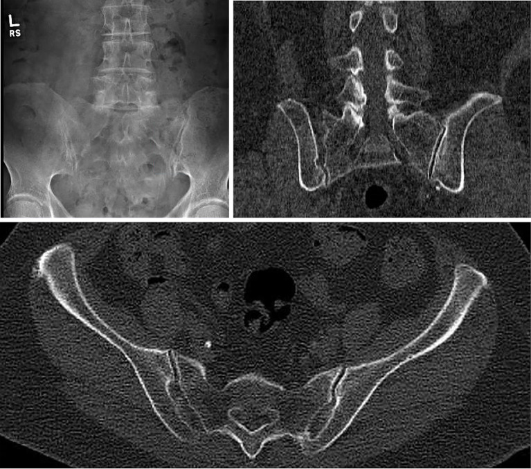
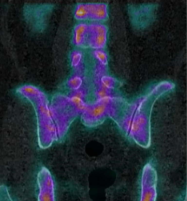
Background: Sacroiliac (SI) joint pain represents a common and often misdiagnosed source of low back and buttock pain. Importantly, SI joint pain can present with lower extremity radicular-type pain and closely mimic a herniated vertebral disc. During the physical exam, nearly all patients with back pain should be evaluated using provocative maneuvers that stress the SI joint. Diagnosis can be further supported with local anesthetic SI joint blocks. Plain radiographs and/or computed tomography (CT) imaging often demonstrate sacroiliac joint degeneration (e.g., joint space narrowing, vacuum phenomenon, osteophyte formation, sclerosis). Other diagnostic studies include magnetic resonance imaging (MRI) of the lumbar spine to evaluate the spinal column, spinal canal, and neural foramina and single photon emission computed tomography scan with CT (SPECT/CT) to identify foci with increased metabolic activity that could be pain generators.
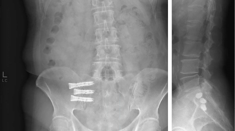
Case description: We present the case of a 76-year-old woman with a 1-year history of progressive left-sided low back pain that progressed along an S1 radicular distribution and became debilitating, interfering with their activities of daily living. Specifically, the pain radiated along the left buttocks, down the posterior aspect of the leg, and into the heel. SPECT/CT demonstrated non-specific, symmetric radiotracer uptake within the bilateral SI joints. She had a positive response to two SI joint injections. The patient was ultimately treated with a SI joint fusion. This case raises questions regarding the sensitivity and specificity of SPECT/CT for SI joint pain. A minimally invasive surgery (MIS) lateral SI joint fusion using navigated and fenestrated screws can provide significant pain relief.
Conclusions: This case illustrates the diverse presentation of SI joint pain, the diagnostic process including the use of SPECT/CT, and the application of MIS fusion techniques for treatment.

 求助内容:
求助内容: 应助结果提醒方式:


