Enhancing Specificity in Predicting Axillary Lymph Node Metastasis in Breast Cancer through an Interpretable Machine Learning Model with CEM and Ultrasound Integration.
{"title":"Enhancing Specificity in Predicting Axillary Lymph Node Metastasis in Breast Cancer through an Interpretable Machine Learning Model with CEM and Ultrasound Integration.","authors":"Weimin Xu, Bowen Zheng, Chanjuan Wen, Hui Zeng, Sina Wang, Zilong He, Xin Liao, Weiguo Chen, Yingjia Li, Genggeng Qin","doi":"10.1177/15330338251334735","DOIUrl":null,"url":null,"abstract":"<p><p>IntroductionThe study aims to evaluate the performance of an interpretable machine learning model in predicting preoperative axillary lymph node metastasis using primary breast cancer and lymph node features derived from contrast-enhanced mammography (CEM) and ultrasound (US) breast imaging reporting and data systems (BI-RADS).MethodsThis retrospective study included patients diagnosed with primary breast cancer. Two experienced radiologists extracted the BI-RADS features from the largest cross-section of the lesions and axillary lymph nodes based on CEM and US images, creating three datasets. Each dataset will train six base models to predict axillary lymph nodes, with pathological results serving as the gold standard. The top three models were used to train the five ensemble models. Additionally, SHapley Additive exPlanations (SHAP) was used to interpret the optimal model. The receiver-operating characteristic curve (ROC) and AUC were used to evaluate model performance.ResultsThis study involved 292 female patients, of whom 99 had axillary lymph node metastasis and 193 did not. The combination of CEM and ultrasound BI-RADS demonstrated the best performance in predicting axillary lymph node metastasis. Among these, the LightGBM achieved the highest AUC (0.762) and specificity (86.67%, while the ensemble model using RF as the meta-model had an AUC (0.754) and specificity (83.33%. The most important variables identified by SHAP were the long diameters of the lymph nodes in the CEM recombined image, along with their complete morphology in the low-energy image.ConclusionThe machine learning model using CEM and US BI-RADS features accurately predicted axillary lymph node metastasis before surgery, thereby serving as a valuable tool for clinical decision-making in patients with breast cancer.</p>","PeriodicalId":22203,"journal":{"name":"Technology in Cancer Research & Treatment","volume":"24 ","pages":"15330338251334735"},"PeriodicalIF":2.8000,"publicationDate":"2025-01-01","publicationTypes":"Journal Article","fieldsOfStudy":null,"isOpenAccess":false,"openAccessPdf":"https://www.ncbi.nlm.nih.gov/pmc/articles/PMC12035205/pdf/","citationCount":"0","resultStr":null,"platform":"Semanticscholar","paperid":null,"PeriodicalName":"Technology in Cancer Research & Treatment","FirstCategoryId":"3","ListUrlMain":"https://doi.org/10.1177/15330338251334735","RegionNum":4,"RegionCategory":"医学","ArticlePicture":[],"TitleCN":null,"AbstractTextCN":null,"PMCID":null,"EPubDate":"2025/4/17 0:00:00","PubModel":"Epub","JCR":"Q3","JCRName":"ONCOLOGY","Score":null,"Total":0}
引用次数: 0
Abstract
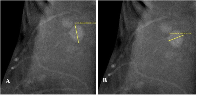
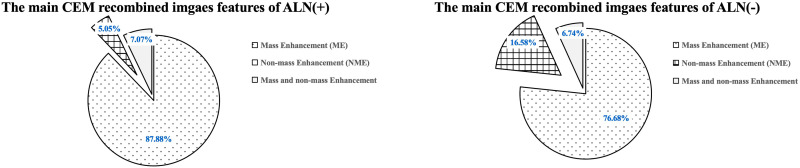
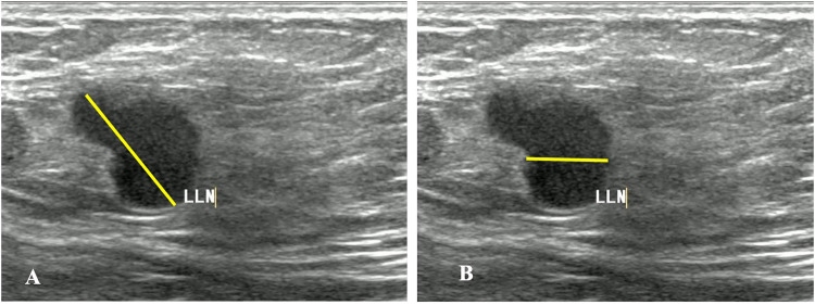
IntroductionThe study aims to evaluate the performance of an interpretable machine learning model in predicting preoperative axillary lymph node metastasis using primary breast cancer and lymph node features derived from contrast-enhanced mammography (CEM) and ultrasound (US) breast imaging reporting and data systems (BI-RADS).MethodsThis retrospective study included patients diagnosed with primary breast cancer. Two experienced radiologists extracted the BI-RADS features from the largest cross-section of the lesions and axillary lymph nodes based on CEM and US images, creating three datasets. Each dataset will train six base models to predict axillary lymph nodes, with pathological results serving as the gold standard. The top three models were used to train the five ensemble models. Additionally, SHapley Additive exPlanations (SHAP) was used to interpret the optimal model. The receiver-operating characteristic curve (ROC) and AUC were used to evaluate model performance.ResultsThis study involved 292 female patients, of whom 99 had axillary lymph node metastasis and 193 did not. The combination of CEM and ultrasound BI-RADS demonstrated the best performance in predicting axillary lymph node metastasis. Among these, the LightGBM achieved the highest AUC (0.762) and specificity (86.67%, while the ensemble model using RF as the meta-model had an AUC (0.754) and specificity (83.33%. The most important variables identified by SHAP were the long diameters of the lymph nodes in the CEM recombined image, along with their complete morphology in the low-energy image.ConclusionThe machine learning model using CEM and US BI-RADS features accurately predicted axillary lymph node metastasis before surgery, thereby serving as a valuable tool for clinical decision-making in patients with breast cancer.
期刊介绍:
Technology in Cancer Research & Treatment (TCRT) is a JCR-ranked, broad-spectrum, open access, peer-reviewed publication whose aim is to provide researchers and clinicians with a platform to share and discuss developments in the prevention, diagnosis, treatment, and monitoring of cancer.
 求助内容:
求助内容: 应助结果提醒方式:


