Njenga R Kamau, Michelle R Tamplin, Chu-Yu Lee, Eric D Axelson, Isabella M Grumbach, Michael S Petronek
{"title":"Combined MR Volumetry and T2* Relaxometry Reveals the Olfactory System as an Iron-Dependent Structure Affected by Radiation.","authors":"Njenga R Kamau, Michelle R Tamplin, Chu-Yu Lee, Eric D Axelson, Isabella M Grumbach, Michael S Petronek","doi":"10.3390/neurolint17040053","DOIUrl":null,"url":null,"abstract":"<p><p><b>Background/Objectives:</b> Radiation therapy can often lead to structural and functional changes in the brain resulting in radiation-induced brain injury. This study investigates the MRI-detectable effects of whole-brain irradiation across all neuroanatomical structures in adult mice, with a specific focus on T2* MRI measurements, to evaluate regions that may be particularly sensitive to iron accumulation. <b>Methods:</b> One year following irradiation or sham treatment, mice were imaged with a 7T MRI to evaluate changes in regional volume and T2* relaxation times across more than 652 neuroanatomical using the DSURQE mouse brain atlas. <b>Results:</b> Statistical analysis identified 301 altered regions with respect to regional volume and 85 regions with respect to T2* relaxation showing significant differences relative to the control group (<i>p</i> < 0.05). Further data refinement, including the consolidation of redundant, bi-lateral structures revealed 18 subregions with significant changes in both volume and T2*. The data refinement revealed that the most represented system was the olfactory system (8/18 regions, 44%). The olfactory regions also showed the most pronounced changes and greatest correlation between the two metrics. <b>Conclusions:</b> These findings are suggestive that ionizing radiation may cause a pronounced disruption in the olfactory system that coincides with potential iron accumulation.</p>","PeriodicalId":19130,"journal":{"name":"Neurology International","volume":"17 4","pages":""},"PeriodicalIF":3.0000,"publicationDate":"2025-04-08","publicationTypes":"Journal Article","fieldsOfStudy":null,"isOpenAccess":false,"openAccessPdf":"https://www.ncbi.nlm.nih.gov/pmc/articles/PMC12029731/pdf/","citationCount":"0","resultStr":null,"platform":"Semanticscholar","paperid":null,"PeriodicalName":"Neurology International","FirstCategoryId":"1085","ListUrlMain":"https://doi.org/10.3390/neurolint17040053","RegionNum":0,"RegionCategory":null,"ArticlePicture":[],"TitleCN":null,"AbstractTextCN":null,"PMCID":null,"EPubDate":"","PubModel":"","JCR":"Q2","JCRName":"CLINICAL NEUROLOGY","Score":null,"Total":0}
引用次数: 0
Abstract
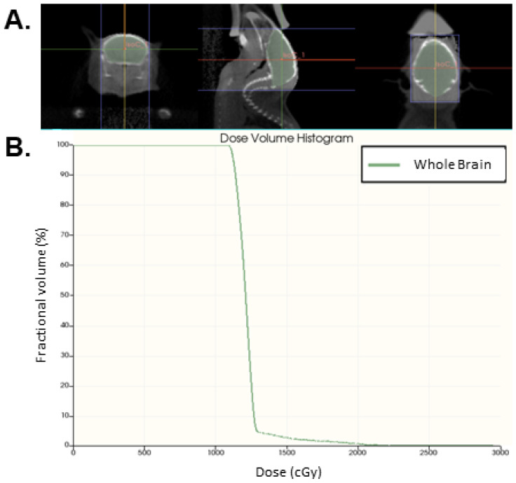
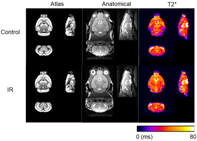
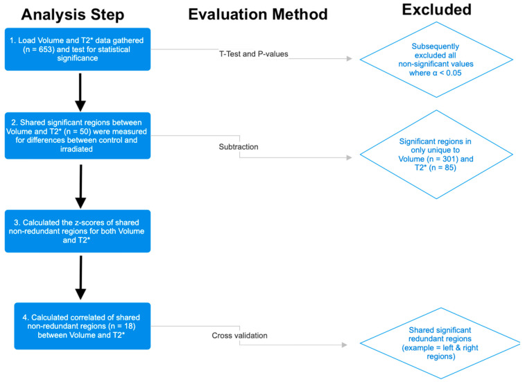
Background/Objectives: Radiation therapy can often lead to structural and functional changes in the brain resulting in radiation-induced brain injury. This study investigates the MRI-detectable effects of whole-brain irradiation across all neuroanatomical structures in adult mice, with a specific focus on T2* MRI measurements, to evaluate regions that may be particularly sensitive to iron accumulation. Methods: One year following irradiation or sham treatment, mice were imaged with a 7T MRI to evaluate changes in regional volume and T2* relaxation times across more than 652 neuroanatomical using the DSURQE mouse brain atlas. Results: Statistical analysis identified 301 altered regions with respect to regional volume and 85 regions with respect to T2* relaxation showing significant differences relative to the control group (p < 0.05). Further data refinement, including the consolidation of redundant, bi-lateral structures revealed 18 subregions with significant changes in both volume and T2*. The data refinement revealed that the most represented system was the olfactory system (8/18 regions, 44%). The olfactory regions also showed the most pronounced changes and greatest correlation between the two metrics. Conclusions: These findings are suggestive that ionizing radiation may cause a pronounced disruption in the olfactory system that coincides with potential iron accumulation.

 求助内容:
求助内容: 应助结果提醒方式:


