Temporal Shift When Comparing Contrast-Agent Concentration Curves Estimated Using Quantitative Susceptibility Mapping (QSM) and ΔR2*: The Association Between Vortex Parameters and Oxygen Extraction Fraction.
IF 2.2 4区 医学Q2 RADIOLOGY, NUCLEAR MEDICINE & MEDICAL IMAGING
Ronnie Wirestam, Anna Lundberg, Linda Knutsson, Emelie Lind
{"title":"Temporal Shift When Comparing Contrast-Agent Concentration Curves Estimated Using Quantitative Susceptibility Mapping (QSM) and ΔR2*: The Association Between Vortex Parameters and Oxygen Extraction Fraction.","authors":"Ronnie Wirestam, Anna Lundberg, Linda Knutsson, Emelie Lind","doi":"10.3390/tomography11040046","DOIUrl":null,"url":null,"abstract":"<p><strong>Background: </strong>When plotting data points corresponding to the contrast-agent-induced change in transverse relaxation rate from a dynamic gradient-echo (GRE) magnetic resonance imaging (MRI) study versus a corresponding spin-echo study, a loop or vortex curve rather than a reversible line is formed. The vortex curve area is likely to reflect vessel architecture and oxygenation level. In this study, the vortex effect seen when using only GRE-based estimates, i.e., contrast-agent concentration based on GRE transverse relaxation rate and contrast-agent concentration based on quantitative susceptibility mapping (QSM), was investigated.</p><p><strong>Methods: </strong>Twenty healthy volunteers were examined using 3 T MRI. Magnitude and phase dynamic contrast-enhanced MRI (DSC-MRI) data were obtained using GRE echo-planar imaging. Vortex curves for grey-matter (GM) regions and for arterial input function (AIF) data were constructed by plotting concentration based on GRE transverse relaxation rate versus concentration based on QSM. Vortex parameters (vortex area and normalised vortex width) were compared with QSM-based whole-brain OEF estimates obtained using 3D GRE.</p><p><strong>Results: </strong>An obvious vortex effect was observed, and both GM vortex parameters showed a moderate and significant correlation with OEF (r = -0.51, <i>p</i> = 0.02). The vortex parameters for AIF data showed no significant correlation with OEF.</p><p><strong>Conclusions: </strong>GRE-based GM vortex parameters correlated significantly with whole-brain OEF. In agreement with expectations, the corresponding AIF data, representing high fractions of arterial blood, showed no significant correlation. Novel parameters, based solely on standard GRE protocols, are of relevance to investigate, considering that GRE-based DSC-MRI is very common in brain tumour applications.</p>","PeriodicalId":51330,"journal":{"name":"Tomography","volume":"11 4","pages":""},"PeriodicalIF":2.2000,"publicationDate":"2025-04-09","publicationTypes":"Journal Article","fieldsOfStudy":null,"isOpenAccess":false,"openAccessPdf":"https://www.ncbi.nlm.nih.gov/pmc/articles/PMC12031548/pdf/","citationCount":"0","resultStr":null,"platform":"Semanticscholar","paperid":null,"PeriodicalName":"Tomography","FirstCategoryId":"3","ListUrlMain":"https://doi.org/10.3390/tomography11040046","RegionNum":4,"RegionCategory":"医学","ArticlePicture":[],"TitleCN":null,"AbstractTextCN":null,"PMCID":null,"EPubDate":"","PubModel":"","JCR":"Q2","JCRName":"RADIOLOGY, NUCLEAR MEDICINE & MEDICAL IMAGING","Score":null,"Total":0}
引用次数: 0
Abstract
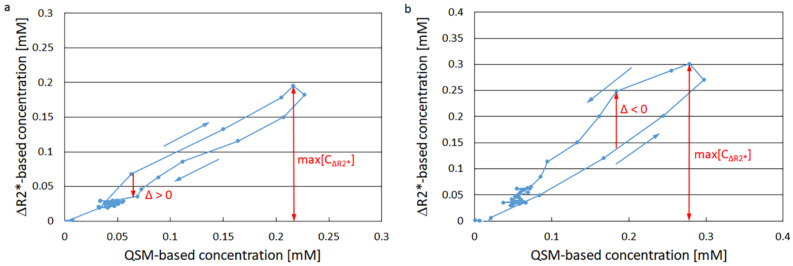
Background: When plotting data points corresponding to the contrast-agent-induced change in transverse relaxation rate from a dynamic gradient-echo (GRE) magnetic resonance imaging (MRI) study versus a corresponding spin-echo study, a loop or vortex curve rather than a reversible line is formed. The vortex curve area is likely to reflect vessel architecture and oxygenation level. In this study, the vortex effect seen when using only GRE-based estimates, i.e., contrast-agent concentration based on GRE transverse relaxation rate and contrast-agent concentration based on quantitative susceptibility mapping (QSM), was investigated.
Methods: Twenty healthy volunteers were examined using 3 T MRI. Magnitude and phase dynamic contrast-enhanced MRI (DSC-MRI) data were obtained using GRE echo-planar imaging. Vortex curves for grey-matter (GM) regions and for arterial input function (AIF) data were constructed by plotting concentration based on GRE transverse relaxation rate versus concentration based on QSM. Vortex parameters (vortex area and normalised vortex width) were compared with QSM-based whole-brain OEF estimates obtained using 3D GRE.
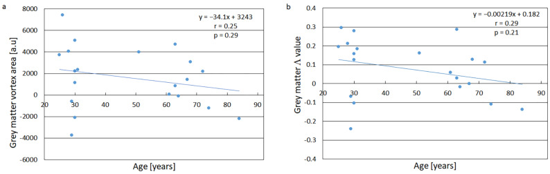
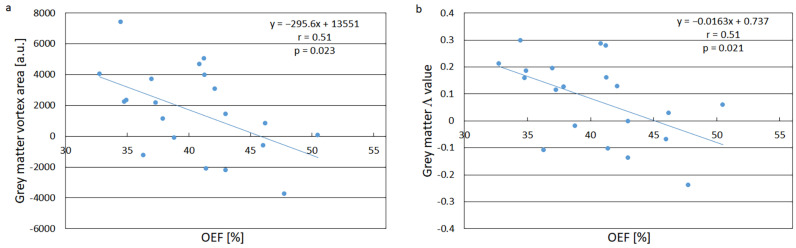
Results: An obvious vortex effect was observed, and both GM vortex parameters showed a moderate and significant correlation with OEF (r = -0.51, p = 0.02). The vortex parameters for AIF data showed no significant correlation with OEF.
Conclusions: GRE-based GM vortex parameters correlated significantly with whole-brain OEF. In agreement with expectations, the corresponding AIF data, representing high fractions of arterial blood, showed no significant correlation. Novel parameters, based solely on standard GRE protocols, are of relevance to investigate, considering that GRE-based DSC-MRI is very common in brain tumour applications.
TomographyMedicine-Radiology, Nuclear Medicine and Imaging
CiteScore
2.70
自引率
10.50%
发文量
222
期刊介绍:
TomographyTM publishes basic (technical and pre-clinical) and clinical scientific articles which involve the advancement of imaging technologies. Tomography encompasses studies that use single or multiple imaging modalities including for example CT, US, PET, SPECT, MR and hyperpolarization technologies, as well as optical modalities (i.e. bioluminescence, photoacoustic, endomicroscopy, fiber optic imaging and optical computed tomography) in basic sciences, engineering, preclinical and clinical medicine.
Tomography also welcomes studies involving exploration and refinement of contrast mechanisms and image-derived metrics within and across modalities toward the development of novel imaging probes for image-based feedback and intervention. The use of imaging in biology and medicine provides unparalleled opportunities to noninvasively interrogate tissues to obtain real-time dynamic and quantitative information required for diagnosis and response to interventions and to follow evolving pathological conditions. As multi-modal studies and the complexities of imaging technologies themselves are ever increasing to provide advanced information to scientists and clinicians.
Tomography provides a unique publication venue allowing investigators the opportunity to more precisely communicate integrated findings related to the diverse and heterogeneous features associated with underlying anatomical, physiological, functional, metabolic and molecular genetic activities of normal and diseased tissue. Thus Tomography publishes peer-reviewed articles which involve the broad use of imaging of any tissue and disease type including both preclinical and clinical investigations. In addition, hardware/software along with chemical and molecular probe advances are welcome as they are deemed to significantly contribute towards the long-term goal of improving the overall impact of imaging on scientific and clinical discovery.

 求助内容:
求助内容: 应助结果提醒方式:


