Dental pulp lymphatic vessel dynamics during tooth development and pulp stimulation in rodents
Abstract
Aim
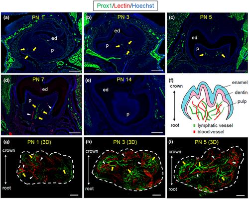
The anatomy and functions of lymphatic vessels (LV) in mammals remain poorly understood compared to the blood vascular system. In particular, whether or not LV exist in the dental pulp is still controversial. This study aims to identify the existence of LV in the mouse dental pulp using a Prox1 (Prospero homeobox 1)-eGFP transgenic mouse model combined with a tissue-clearing technique.
Methodology
Mandible or mandibular first molars of Prox1-eGFP mice were extracted, cleared for whole-mount observation and imaged using confocal microscopy. Dylight 594-lectin was injected intracardially to differentiate Prox1-eGFP+ LV from the blood vascular network. To further determine if pulpal LV act as an interstitial fluid drainage system, we examined ink absorption in surgically exposed dental pulp of mandibular first molars.
Results
At the early stage of tooth development, abundant Prox1-eGFP+ LV distinct from lectin-labelled blood vessels were present in a dental pulp. However, after the initiation of root development, the expression of Prox1-eGFP in dental pulp decreased. In mature dental pulp, Prox1-eGFP+ LV was scattered with a discontinuous lumen. In response to a non-infectious transient pulp stimulation (TPS), the Prox1-eGFP+ LV increased in number and diameter with continuous lumen reaching the apical foramen. Ink particles applied to exposed dental pulp are distributed throughout the dental pulp via interstitial spaces and vessel-like structures. Histological evaluation revealed that ink particles were mainly present in the cell-free zone. However, due to TPS, ink particles were taken up into Prox1-eGFP+ LV.
Conclusion
Our findings suggest the presence of LV in the mature dental pulp that contributes to fluid drainage in this tissue together with the extravascular pathway.

 求助内容:
求助内容: 应助结果提醒方式:


