Is two-dimensional oblique parasagittal ultrasound imaging valid for levator ani muscle assessment?

IF 6.1 1区 医学 Q1 ACOUSTICS
K L Shek, H P Dietz
{"title":"Is two-dimensional oblique parasagittal ultrasound imaging valid for levator ani muscle assessment?","authors":"K L Shek, H P Dietz","doi":"10.1002/uog.29236","DOIUrl":null,"url":null,"abstract":"<p><strong>Objective: </strong>To evaluate the validity of two-dimensional (2D) oblique parasagittal ultrasound imaging to assess levator ani muscle avulsion.</p><p><strong>Methods: </strong>This was a cross-sectional prospective study of women attending a tertiary urogynecological service between February 2021 and August 2022. All women underwent a standardized interview, pelvic organ prolapse quantification (POP-Q) assessment and four-dimensional transperineal ultrasound. 2D oblique parasagittal ultrasound imaging was performed by rotating the transducer 10-20° from the midline to line up the main transducer axis with the fiber direction of the puborectalis muscle, followed by a full parasagittal sweep of the hiatus at rest. Postprocessing of archived ultrasound volume data was performed at a later date, blinded to all other data. Findings were compared with levator ani assessment results obtained previously using three-dimensional tomographic ultrasound imaging (TUI). Diagnosis of levator ani avulsion on TUI and oblique parasagittal imaging was analyzed for associations with pelvic organ prolapse (POP).</p><p><strong>Results: </strong>The datasets of 484 women were analyzed. Mean age was 58 (range, 16-94) years, mean body mass index was 30 (range, 17-65) kg/m<sup>2</sup> and mean parity was 2.6 (range, 0-8). POP symptoms were reported by 278 (57%) women. Clinically and sonographically significant POP was found in 385 (80%) and 350 (72%) women, respectively. Levator ani avulsion was diagnosed in 77 (16%) women on TUI and in 90 (18.6%) women on oblique parasagittal ultrasound imaging, with fair agreement between the two methods (Cohen's kappa of 0.365). There were significant associations between levator ani avulsion on 2D ultrasound imaging and POP diagnosis on clinical examination (odds ratio (OR), 2.88 (95% CI, 1.34-6.18); P = 0.005) and on ultrasound (OR, 2.92 (95% CI, 1.53-5.55); P = 0.001), but these associations were much stronger for TUI (P < 0.001 for both).</p><p><strong>Conclusion: </strong>There was limited agreement between tomographic and oblique parasagittal ultrasound diagnosis of levator ani muscle avulsion. The latter technique has some validity for levator ani assessment but is clearly less valid than TUI. © 2025 The Author(s). Ultrasound in Obstetrics & Gynecology published by John Wiley & Sons Ltd on behalf of International Society of Ultrasound in Obstetrics and Gynecology.</p>","PeriodicalId":23454,"journal":{"name":"Ultrasound in Obstetrics & Gynecology","volume":" ","pages":""},"PeriodicalIF":6.1000,"publicationDate":"2025-05-13","publicationTypes":"Journal Article","fieldsOfStudy":null,"isOpenAccess":false,"openAccessPdf":"","citationCount":"0","resultStr":null,"platform":"Semanticscholar","paperid":null,"PeriodicalName":"Ultrasound in Obstetrics & Gynecology","FirstCategoryId":"3","ListUrlMain":"https://doi.org/10.1002/uog.29236","RegionNum":1,"RegionCategory":"医学","ArticlePicture":[],"TitleCN":null,"AbstractTextCN":null,"PMCID":null,"EPubDate":"","PubModel":"","JCR":"Q1","JCRName":"ACOUSTICS","Score":null,"Total":0}
引用次数: 0

Abstract

Objective: To evaluate the validity of two-dimensional (2D) oblique parasagittal ultrasound imaging to assess levator ani muscle avulsion.

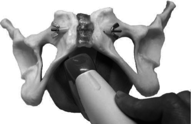
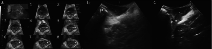
Methods: This was a cross-sectional prospective study of women attending a tertiary urogynecological service between February 2021 and August 2022. All women underwent a standardized interview, pelvic organ prolapse quantification (POP-Q) assessment and four-dimensional transperineal ultrasound. 2D oblique parasagittal ultrasound imaging was performed by rotating the transducer 10-20° from the midline to line up the main transducer axis with the fiber direction of the puborectalis muscle, followed by a full parasagittal sweep of the hiatus at rest. Postprocessing of archived ultrasound volume data was performed at a later date, blinded to all other data. Findings were compared with levator ani assessment results obtained previously using three-dimensional tomographic ultrasound imaging (TUI). Diagnosis of levator ani avulsion on TUI and oblique parasagittal imaging was analyzed for associations with pelvic organ prolapse (POP).

Results: The datasets of 484 women were analyzed. Mean age was 58 (range, 16-94) years, mean body mass index was 30 (range, 17-65) kg/m2 and mean parity was 2.6 (range, 0-8). POP symptoms were reported by 278 (57%) women. Clinically and sonographically significant POP was found in 385 (80%) and 350 (72%) women, respectively. Levator ani avulsion was diagnosed in 77 (16%) women on TUI and in 90 (18.6%) women on oblique parasagittal ultrasound imaging, with fair agreement between the two methods (Cohen's kappa of 0.365). There were significant associations between levator ani avulsion on 2D ultrasound imaging and POP diagnosis on clinical examination (odds ratio (OR), 2.88 (95% CI, 1.34-6.18); P = 0.005) and on ultrasound (OR, 2.92 (95% CI, 1.53-5.55); P = 0.001), but these associations were much stronger for TUI (P < 0.001 for both).

Conclusion: There was limited agreement between tomographic and oblique parasagittal ultrasound diagnosis of levator ani muscle avulsion. The latter technique has some validity for levator ani assessment but is clearly less valid than TUI. © 2025 The Author(s). Ultrasound in Obstetrics & Gynecology published by John Wiley & Sons Ltd on behalf of International Society of Ultrasound in Obstetrics and Gynecology.

二维斜矢状旁超声成像对提肛肌评估有效吗?
目的:评价二维斜矢旁超声成像评价提肛肌撕脱伤的有效性。方法:这是一项横断面前瞻性研究,研究对象是2021年2月至2022年8月期间在第三泌尿妇科就诊的女性。所有女性均接受标准化访谈、盆腔器官脱垂定量(POP-Q)评估和四维会阴超声检查。将换能器从中线旋转10-20°,使换能器主轴与耻骨直肠肌纤维方向对齐,进行二维斜矢状旁超声成像,然后对静息间隙进行全斜矢状旁扫描。存档的超声体积数据的后处理在稍后的日期进行,对所有其他数据视而不见。结果与先前使用三维层析超声成像(TUI)获得的提肛肌评估结果进行了比较。分析了提肛肌撕脱脱与盆腔器官脱垂(POP)的相关性。结果:分析了484例妇女的数据集。平均年龄58岁(范围16 ~ 94岁),平均体重指数30(范围17 ~ 65)kg/m2,平均胎次2.6(范围0 ~ 8)。278名(57%)女性报告了POP症状。临床和超声检查分别有385例(80%)和350例(72%)的女性发现明显的POP。77例(16%)女性在TUI检查中被诊断为提肛肌撕脱,90例(18.6%)女性在斜矢状旁超声检查中被诊断为提肛肌撕脱,两种方法之间基本一致(Cohen’s kappa为0.365)。二维超声图像上的提肛肌撕脱伤与临床检查中的POP诊断有显著相关性(优势比(OR), 2.88 (95% CI, 1.34-6.18);P = 0.005)和超声检查(OR, 2.92 (95% CI, 1.53-5.55);P = 0.001),但这些相关性在TUI中更强(P结论:提肛肌撕脱的断层扫描和斜矢状旁超声诊断的一致性有限。后一种方法对提肛肌的评估有一定的有效性,但明显不如TUI有效。©2025作者。妇产科学超声由John Wiley & Sons Ltd代表国际妇产科学超声学会出版。
本文章由计算机程序翻译,如有差异,请以英文原文为准。
求助全文
约1分钟内获得全文 求助全文
来源期刊
CiteScore
12.30
自引率
14.10%
发文量
891
审稿时长
1 months
期刊介绍: Ultrasound in Obstetrics & Gynecology (UOG) is the official journal of the International Society of Ultrasound in Obstetrics and Gynecology (ISUOG) and is considered the foremost international peer-reviewed journal in the field. It publishes cutting-edge research that is highly relevant to clinical practice, which includes guidelines, expert commentaries, consensus statements, original articles, and systematic reviews. UOG is widely recognized and included in prominent abstract and indexing databases such as Index Medicus and Current Contents.
×
引用
GB/T 7714-2015
复制
MLA
复制
APA
复制
导出至
BibTeX EndNote RefMan NoteFirst NoteExpress
×
提示
您的信息不完整,为了账户安全,请先补充。
现在去补充
×
提示
您因"违规操作"
具体请查看互助需知
我知道了
×
提示
确定
请完成安全验证×
copy
已复制链接
快去分享给好友吧!
我知道了
右上角分享
点击右上角分享
0
联系我们:info@booksci.cn Book学术提供免费学术资源搜索服务,方便国内外学者检索中英文文献。致力于提供最便捷和优质的服务体验。 Copyright © 2023 布克学术 All rights reserved.
京ICP备2023020795号-1
ghs 京公网安备 11010802042870号
Book学术文献互助
Book学术文献互助群
群 号:481959085
Book学术官方微信