Aksel Laudon, Zhaoze Wang, Anqi Zou, Richa Sharma, Jiayi Ji, Winston Tan, Connor Kim, Yingzhe Qian, Qin Ye, Hui Chen, Joel M Henderson, Chao Zhang, Vijaya B Kolachalama, Weining Lu
{"title":"Digital pathology assessment of kidney glomerular filtration barrier ultrastructure in an animal model of podocytopathy.","authors":"Aksel Laudon, Zhaoze Wang, Anqi Zou, Richa Sharma, Jiayi Ji, Winston Tan, Connor Kim, Yingzhe Qian, Qin Ye, Hui Chen, Joel M Henderson, Chao Zhang, Vijaya B Kolachalama, Weining Lu","doi":"10.1093/biomethods/bpaf024","DOIUrl":null,"url":null,"abstract":"<p><p>Transmission electron microscopy (TEM) images can visualize kidney glomerular filtration barrier ultrastructure, including the glomerular basement membrane (GBM) and podocyte foot processes (PFP). Podocytopathy is associated with glomerular filtration barrier morphological changes observed experimentally and clinically by measuring GBM or PFP width. However, these measurements are currently performed manually. This limits research on podocytopathy disease mechanisms and therapeutics due to labor intensiveness and inter-operator variability. We developed a deep learning-based digital pathology computational method to measure GBM and PFP width in TEM images from the kidneys of Integrin-Linked Kinase (ILK) podocyte-specific conditional knockout (cKO) mouse, an animal model of podocytopathy, compared to wild-type (WT) control mouse. We obtained TEM images from WT and ILK cKO littermate mice at 4 weeks old. Our automated method was composed of two stages: a U-Net model for GBM segmentation, followed by an image processing algorithm for GBM and PFP width measurement. We evaluated its performance with a 4-fold cross-validation study on WT and ILK cKO mouse kidney pairs. Mean [95% confidence interval (CI)] GBM segmentation accuracy, calculated as Jaccard index, was 0.73 (0.70-0.76) for WT and 0.85 (0.83-0.87) for ILK cKO TEM images. Automated and manual GBM width measurements were similar for both WT (<i>P</i> = .49) and ILK cKO (<i>P</i> = .06) specimens. While automated and manual PFP width measurements were similar for WT (<i>P</i> = .89), they differed for ILK cKO (<i>P</i> < .05) specimens. WT and ILK cKO specimens were morphologically distinguishable by manual GBM (<i>P</i> < .05) and PFP (<i>P</i> < .05) width measurements. This phenotypic difference was reflected in the automated GBM (<i>P</i> < .05) more than PFP (<i>P</i> = .06) widths. Our deep learning-based digital pathology tool automated measurements in a mouse model of podocytopathy. This proposed method provides high-throughput, objective morphological analysis and could facilitate podocytopathy research.</p>","PeriodicalId":36528,"journal":{"name":"Biology Methods and Protocols","volume":"10 1","pages":"bpaf024"},"PeriodicalIF":1.3000,"publicationDate":"2025-03-28","publicationTypes":"Journal Article","fieldsOfStudy":null,"isOpenAccess":false,"openAccessPdf":"https://www.ncbi.nlm.nih.gov/pmc/articles/PMC11992336/pdf/","citationCount":"0","resultStr":null,"platform":"Semanticscholar","paperid":null,"PeriodicalName":"Biology Methods and Protocols","FirstCategoryId":"1085","ListUrlMain":"https://doi.org/10.1093/biomethods/bpaf024","RegionNum":0,"RegionCategory":null,"ArticlePicture":[],"TitleCN":null,"AbstractTextCN":null,"PMCID":null,"EPubDate":"2025/1/1 0:00:00","PubModel":"eCollection","JCR":"Q3","JCRName":"BIOCHEMICAL RESEARCH METHODS","Score":null,"Total":0}
引用次数: 0
Abstract
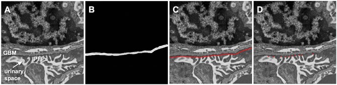
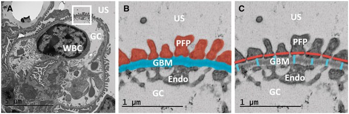
Transmission electron microscopy (TEM) images can visualize kidney glomerular filtration barrier ultrastructure, including the glomerular basement membrane (GBM) and podocyte foot processes (PFP). Podocytopathy is associated with glomerular filtration barrier morphological changes observed experimentally and clinically by measuring GBM or PFP width. However, these measurements are currently performed manually. This limits research on podocytopathy disease mechanisms and therapeutics due to labor intensiveness and inter-operator variability. We developed a deep learning-based digital pathology computational method to measure GBM and PFP width in TEM images from the kidneys of Integrin-Linked Kinase (ILK) podocyte-specific conditional knockout (cKO) mouse, an animal model of podocytopathy, compared to wild-type (WT) control mouse. We obtained TEM images from WT and ILK cKO littermate mice at 4 weeks old. Our automated method was composed of two stages: a U-Net model for GBM segmentation, followed by an image processing algorithm for GBM and PFP width measurement. We evaluated its performance with a 4-fold cross-validation study on WT and ILK cKO mouse kidney pairs. Mean [95% confidence interval (CI)] GBM segmentation accuracy, calculated as Jaccard index, was 0.73 (0.70-0.76) for WT and 0.85 (0.83-0.87) for ILK cKO TEM images. Automated and manual GBM width measurements were similar for both WT (P = .49) and ILK cKO (P = .06) specimens. While automated and manual PFP width measurements were similar for WT (P = .89), they differed for ILK cKO (P < .05) specimens. WT and ILK cKO specimens were morphologically distinguishable by manual GBM (P < .05) and PFP (P < .05) width measurements. This phenotypic difference was reflected in the automated GBM (P < .05) more than PFP (P = .06) widths. Our deep learning-based digital pathology tool automated measurements in a mouse model of podocytopathy. This proposed method provides high-throughput, objective morphological analysis and could facilitate podocytopathy research.
透射电子显微镜(TEM)图像可以显示肾小球滤过屏障的超微结构,包括肾小球基底膜(GBM)和足细胞足突(PFP)。足细胞病与肾小球滤过屏障形态学改变有关,通过实验和临床测量GBM或PFP宽度。然而,这些测量目前是手动执行的。由于劳动强度和操作者之间的差异,这限制了足细胞病疾病机制和治疗方法的研究。我们开发了一种基于深度学习的数字病理学计算方法,用于测量整合素连接激酶(ILK)足细胞特异性条件敲除(cKO)小鼠肾脏TEM图像中的GBM和PFP宽度,这是一种足细胞病变动物模型,与野生型(WT)对照小鼠进行比较。我们在4周龄时获得了WT和ILK cKO同窝小鼠的TEM图像。我们的自动化方法由两个阶段组成:用于GBM分割的U-Net模型,然后是用于GBM和PFP宽度测量的图像处理算法。我们通过对WT和ILK cKO小鼠肾对的4倍交叉验证研究来评估其性能。平均[95%置信区间(CI)] GBM分割精度,以Jaccard指数计算,WT为0.73 (0.70-0.76),ILK cKO TEM图像为0.85(0.83-0.87)。对于WT (P = .49)和ILK cKO (P = .06)标本,自动和手动GBM宽度测量相似。虽然自动和手动PFP宽度测量在WT上相似(P = .89),但在ILK cKO上不同(P P P P P P = .06)。我们基于深度学习的数字病理工具在小鼠足细胞病模型中自动测量。该方法提供了高通量、客观的形态学分析,有助于足细胞病的研究。


 求助内容:
求助内容: 应助结果提醒方式:


