Ajay Chhabra, K P Ramya, B Saravana Prathap, Bhavana Murjani, Priyanka Yadav
{"title":"A Comparative Study of Four Rotary Endodontic File Systems: Assessing Canal Transportation and Centering Ability using Cone-beam Computed Tomography.","authors":"Ajay Chhabra, K P Ramya, B Saravana Prathap, Bhavana Murjani, Priyanka Yadav","doi":"10.4103/ccd.ccd_379_24","DOIUrl":null,"url":null,"abstract":"<p><strong>Aim: </strong>The aim of this study was to compare the canal transportation and centering ability of four rotary endodontic file systems using cone-beam computed tomography (CBCT).</p><p><strong>Methodology: </strong>This was an <i>in vitro</i> study where 40 extracted single-rooted single canal human premolar teeth were used. CBCT scans of all the teeth were taken before instrumentation and were randomly divided into four groups, with 10 samples in each; Group I-Protaper Next (PN), Group II-Protaper Gold (PG), Group III-Neo Endo Flex (NE), and Group IV-Gen Endo (GE). After cleaning and shaping, the canals with respective file systems postinstrumentation scans were performed, and the two scans were compared to determine canal transportation and centering ability at 3, 6, and 9 mm, from the apex.</p><p><strong>Results: </strong>Statistical analysis was performed using the Kruskal-Wallis test where the results showed no statistically significant difference among the tested groups in canal transportation and centering ability at the coronal, middle, and cervical third (<i>P</i> > 0.05).</p><p><strong>Conclusion: </strong>Under the conditions of the <i>in vitro</i> study PN, PG, NE, and GE file systems exhibited comparable behavior in terms of canal transportation and centering ability, observational data suggested that Group 1 (PN) and Group 4 (GE) demonstrated superior centering ability and reduced canal transportation.</p>","PeriodicalId":10632,"journal":{"name":"Contemporary Clinical Dentistry","volume":"16 1","pages":"44-48"},"PeriodicalIF":1.1000,"publicationDate":"2025-01-01","publicationTypes":"Journal Article","fieldsOfStudy":null,"isOpenAccess":false,"openAccessPdf":"https://www.ncbi.nlm.nih.gov/pmc/articles/PMC12014003/pdf/","citationCount":"0","resultStr":null,"platform":"Semanticscholar","paperid":null,"PeriodicalName":"Contemporary Clinical Dentistry","FirstCategoryId":"1085","ListUrlMain":"https://doi.org/10.4103/ccd.ccd_379_24","RegionNum":0,"RegionCategory":null,"ArticlePicture":[],"TitleCN":null,"AbstractTextCN":null,"PMCID":null,"EPubDate":"2025/3/25 0:00:00","PubModel":"Epub","JCR":"Q3","JCRName":"DENTISTRY, ORAL SURGERY & MEDICINE","Score":null,"Total":0}
引用次数: 0
Abstract
Aim: The aim of this study was to compare the canal transportation and centering ability of four rotary endodontic file systems using cone-beam computed tomography (CBCT).
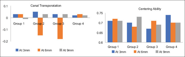
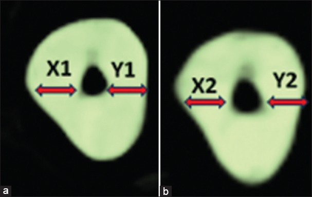
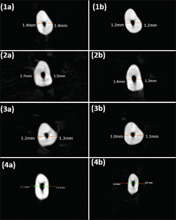
Methodology: This was an in vitro study where 40 extracted single-rooted single canal human premolar teeth were used. CBCT scans of all the teeth were taken before instrumentation and were randomly divided into four groups, with 10 samples in each; Group I-Protaper Next (PN), Group II-Protaper Gold (PG), Group III-Neo Endo Flex (NE), and Group IV-Gen Endo (GE). After cleaning and shaping, the canals with respective file systems postinstrumentation scans were performed, and the two scans were compared to determine canal transportation and centering ability at 3, 6, and 9 mm, from the apex.
Results: Statistical analysis was performed using the Kruskal-Wallis test where the results showed no statistically significant difference among the tested groups in canal transportation and centering ability at the coronal, middle, and cervical third (P > 0.05).
Conclusion: Under the conditions of the in vitro study PN, PG, NE, and GE file systems exhibited comparable behavior in terms of canal transportation and centering ability, observational data suggested that Group 1 (PN) and Group 4 (GE) demonstrated superior centering ability and reduced canal transportation.
期刊介绍:
The journal Contemporary Clinical Dentistry (CCD) (Print ISSN: 0976-237X, E-ISSN:0976- 2361) is peer-reviewed journal published on behalf of Maharishi Markandeshwar University and issues are published quarterly in the last week of March, June, September and December. The Journal publishes Original research papers, clinical studies, case series strictly of clinical interest. Manuscripts are invited from all specialties of Dentistry i.e. Conservative dentistry and Endodontics, Dentofacial orthopedics and Orthodontics, Oral medicine and Radiology, Oral pathology, Oral surgery, Orodental diseases, Pediatric Dentistry, Periodontics, Clinical aspects of Public Health dentistry and Prosthodontics. Review articles are not accepted. Review, if published, will only be by invitation from eminent scholars and academicians of National and International repute in the field of Medical/Dental education.

 求助内容:
求助内容: 应助结果提醒方式:


