Janusz Blasiak, Elzbieta Pawlowska, Hanna Helotera, Maksim Ionov, Marcin Derwich, Kai Kaarniranta
{"title":"Potential of autophagy in subretinal fibrosis in neovascular age-related macular degeneration.","authors":"Janusz Blasiak, Elzbieta Pawlowska, Hanna Helotera, Maksim Ionov, Marcin Derwich, Kai Kaarniranta","doi":"10.1186/s11658-025-00732-8","DOIUrl":null,"url":null,"abstract":"<p><p>Age-related macular degeneration (AMD) is an eye disease that can lead to legal blindness and vision loss. In its advanced stages, it is classified into dry and neovascular AMD. In neovascular AMD, the formation of new blood vessels disrupts the structure of the retina and induces an inflammatory response. Treatment for neovascular AMD involves antibodies and fusion proteins targeting vascular endothelial growth factor A (VEGFA) and its receptors to inhibit neovascularization and slow vision loss. However, a fraction of patients with neovascular AMD do not respond to therapy. Many of these patients exhibit a subretinal fibrotic scar. Thus, retinal fibrosis may contribute to resistance against anti-VEGFA therapy and the cause of irreversible vision loss in neovascular AMD patients. Retinal pigment epithelium cells, choroidal fibroblasts, and retinal glial cells are crucial in the development of the fibrotic scar as they can undergo a mesenchymal transition mediated by transforming growth factor beta and other molecules, leading to their transdifferentiation into myofibroblasts, which are key players in subretinal fibrosis. Autophagy, a process that removes cellular debris and contributes to the pathogenesis of AMD, regardless of its type, may be stimulated by epithelial-mesenchymal transition and later inhibited. The mesenchymal transition of retinal cells and the dysfunction of the extracellular matrix-the two main aspects of fibrotic scar formation-are associated with impaired autophagy. Nonetheless, the causal relationship between autophagy and subretinal fibrosis remains unknown. This narrative/perspective review presents information on neovascular AMD, subretinal fibrosis, and autophagy, arguing that impaired autophagy may be significant for fibrosis-related resistance to anti-VEGFA therapy in neovascular AMD.</p>","PeriodicalId":9688,"journal":{"name":"Cellular & Molecular Biology Letters","volume":"30 1","pages":"54"},"PeriodicalIF":10.2000,"publicationDate":"2025-04-30","publicationTypes":"Journal Article","fieldsOfStudy":null,"isOpenAccess":false,"openAccessPdf":"https://www.ncbi.nlm.nih.gov/pmc/articles/PMC12044759/pdf/","citationCount":"0","resultStr":null,"platform":"Semanticscholar","paperid":null,"PeriodicalName":"Cellular & Molecular Biology Letters","FirstCategoryId":"99","ListUrlMain":"https://doi.org/10.1186/s11658-025-00732-8","RegionNum":1,"RegionCategory":"生物学","ArticlePicture":[],"TitleCN":null,"AbstractTextCN":null,"PMCID":null,"EPubDate":"","PubModel":"","JCR":"Q1","JCRName":"BIOCHEMISTRY & MOLECULAR BIOLOGY","Score":null,"Total":0}
引用次数: 0
Abstract
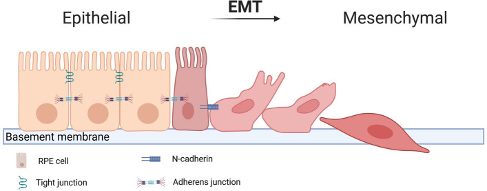
Age-related macular degeneration (AMD) is an eye disease that can lead to legal blindness and vision loss. In its advanced stages, it is classified into dry and neovascular AMD. In neovascular AMD, the formation of new blood vessels disrupts the structure of the retina and induces an inflammatory response. Treatment for neovascular AMD involves antibodies and fusion proteins targeting vascular endothelial growth factor A (VEGFA) and its receptors to inhibit neovascularization and slow vision loss. However, a fraction of patients with neovascular AMD do not respond to therapy. Many of these patients exhibit a subretinal fibrotic scar. Thus, retinal fibrosis may contribute to resistance against anti-VEGFA therapy and the cause of irreversible vision loss in neovascular AMD patients. Retinal pigment epithelium cells, choroidal fibroblasts, and retinal glial cells are crucial in the development of the fibrotic scar as they can undergo a mesenchymal transition mediated by transforming growth factor beta and other molecules, leading to their transdifferentiation into myofibroblasts, which are key players in subretinal fibrosis. Autophagy, a process that removes cellular debris and contributes to the pathogenesis of AMD, regardless of its type, may be stimulated by epithelial-mesenchymal transition and later inhibited. The mesenchymal transition of retinal cells and the dysfunction of the extracellular matrix-the two main aspects of fibrotic scar formation-are associated with impaired autophagy. Nonetheless, the causal relationship between autophagy and subretinal fibrosis remains unknown. This narrative/perspective review presents information on neovascular AMD, subretinal fibrosis, and autophagy, arguing that impaired autophagy may be significant for fibrosis-related resistance to anti-VEGFA therapy in neovascular AMD.
期刊介绍:
Cellular & Molecular Biology Letters is an international journal dedicated to the dissemination of fundamental knowledge in all areas of cellular and molecular biology, cancer cell biology, and certain aspects of biochemistry, biophysics and biotechnology.
 求助内容:
求助内容: 应助结果提醒方式:


