Salma Binzaqr, David Kryza, Anne-Laure Giraudet, Jean Christophe Bernhard, Marine Gross-Goupil, Mokrane Yacoub, Gaelle Margue, Elif Hindié, Clément Morgat
{"title":"Prostate-specific membrane antigen (PSMA) expression in primary and metastatic renal cell cancer (UroCCR-65 study).","authors":"Salma Binzaqr, David Kryza, Anne-Laure Giraudet, Jean Christophe Bernhard, Marine Gross-Goupil, Mokrane Yacoub, Gaelle Margue, Elif Hindié, Clément Morgat","doi":"10.1186/s13550-025-01232-8","DOIUrl":null,"url":null,"abstract":"<p><strong>Background: </strong>Prostate-specific membrane antigen (PSMA) has been shown to be overexpressed in the neo-vasculature of renal cancers. However, studies investigating the pattern of PSMA expression in primary RCC and RCC metastases according to metastatic sites are rare. 44 frozen samples of RCC, 19 primaries (9 clear cell (cc) RCC, 7 papillary (pap) RCC, and 3 chromophobe (ch) RCC) and 25 (24 samples have ccRCC histology and one is unclassified) unpaired metastases (8 from adrenals, 8 from bones, 2 from lungs, 2 from liver and 5 others (1 lymph node, 1 pancreas, 1 brain, 1 gallbladder and 1 muscle)), were available from the UroCCR project (NCT03293563). PSMA expression was assessed by autoradiography using [<sup>177</sup>Lu]Lu-PSMA-617 as binding agent and the specific binding (total binding-non-specific binding) was calculated and expressed as a percentage of total binding. A patient suffering from metastatic ccRCC was also administered [<sup>68</sup>Ga]Ga-PSMA-11 to evaluate PSMA expression.</p><p><strong>Results: </strong>The mean specific binding was 28.9 ± 40.4% for primary renal cancer and 65.0 ± 38.9% for metastasis. Regarding histology, high PSMA expression was depicted in 33.3% of ccRCC, 33.3% of chRCC and 57.1% of papRCC. PSMA was more frequently expressed in primary samples of papRCC histology with renal capsule invasion (p = 0.0286). A higher PSMA-specific binding and a higher number of samples with high PSMA-expression were depicted in metastatic samples. Bone metastasis showed lower binding than other metastatic sites combined (p = 0.0005). The patient suffering from metastatic ccRCC showed high [<sup>68</sup>Ga]Ga-PSMA-11 uptake on known distant metastases and additional site uncovered.</p><p><strong>Conclusion: </strong>PSMA showed high expression in metastases of ccRCC.</p><p><strong>Clinical trial registration: </strong>NCT, NCT03293563, prospectively registered September 20, 2017, http://www.</p><p><strong>Clinicaltrials: </strong>gov .</p>","PeriodicalId":11611,"journal":{"name":"EJNMMI Research","volume":"15 1","pages":"38"},"PeriodicalIF":3.1000,"publicationDate":"2025-04-09","publicationTypes":"Journal Article","fieldsOfStudy":null,"isOpenAccess":false,"openAccessPdf":"https://www.ncbi.nlm.nih.gov/pmc/articles/PMC11981970/pdf/","citationCount":"0","resultStr":null,"platform":"Semanticscholar","paperid":null,"PeriodicalName":"EJNMMI Research","FirstCategoryId":"3","ListUrlMain":"https://doi.org/10.1186/s13550-025-01232-8","RegionNum":3,"RegionCategory":"医学","ArticlePicture":[],"TitleCN":null,"AbstractTextCN":null,"PMCID":null,"EPubDate":"","PubModel":"","JCR":"Q1","JCRName":"RADIOLOGY, NUCLEAR MEDICINE & MEDICAL IMAGING","Score":null,"Total":0}
引用次数: 0
Abstract
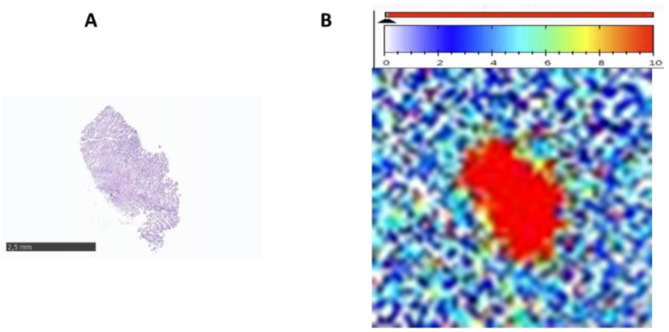
Background: Prostate-specific membrane antigen (PSMA) has been shown to be overexpressed in the neo-vasculature of renal cancers. However, studies investigating the pattern of PSMA expression in primary RCC and RCC metastases according to metastatic sites are rare. 44 frozen samples of RCC, 19 primaries (9 clear cell (cc) RCC, 7 papillary (pap) RCC, and 3 chromophobe (ch) RCC) and 25 (24 samples have ccRCC histology and one is unclassified) unpaired metastases (8 from adrenals, 8 from bones, 2 from lungs, 2 from liver and 5 others (1 lymph node, 1 pancreas, 1 brain, 1 gallbladder and 1 muscle)), were available from the UroCCR project (NCT03293563). PSMA expression was assessed by autoradiography using [177Lu]Lu-PSMA-617 as binding agent and the specific binding (total binding-non-specific binding) was calculated and expressed as a percentage of total binding. A patient suffering from metastatic ccRCC was also administered [68Ga]Ga-PSMA-11 to evaluate PSMA expression.
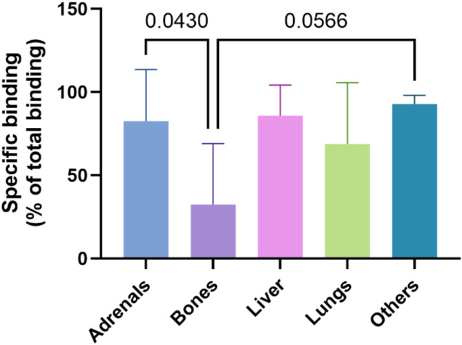
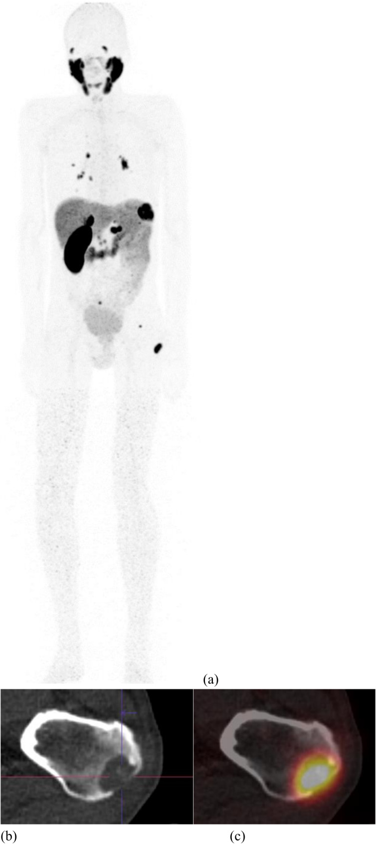
Results: The mean specific binding was 28.9 ± 40.4% for primary renal cancer and 65.0 ± 38.9% for metastasis. Regarding histology, high PSMA expression was depicted in 33.3% of ccRCC, 33.3% of chRCC and 57.1% of papRCC. PSMA was more frequently expressed in primary samples of papRCC histology with renal capsule invasion (p = 0.0286). A higher PSMA-specific binding and a higher number of samples with high PSMA-expression were depicted in metastatic samples. Bone metastasis showed lower binding than other metastatic sites combined (p = 0.0005). The patient suffering from metastatic ccRCC showed high [68Ga]Ga-PSMA-11 uptake on known distant metastases and additional site uncovered.
Conclusion: PSMA showed high expression in metastases of ccRCC.
EJNMMI ResearchRADIOLOGY, NUCLEAR MEDICINE & MEDICAL IMAGING&nb-
CiteScore
5.90
自引率
3.10%
发文量
72
审稿时长
13 weeks
期刊介绍:
EJNMMI Research publishes new basic, translational and clinical research in the field of nuclear medicine and molecular imaging. Regular features include original research articles, rapid communication of preliminary data on innovative research, interesting case reports, editorials, and letters to the editor. Educational articles on basic sciences, fundamental aspects and controversy related to pre-clinical and clinical research or ethical aspects of research are also welcome. Timely reviews provide updates on current applications, issues in imaging research and translational aspects of nuclear medicine and molecular imaging technologies.
The main emphasis is placed on the development of targeted imaging with radiopharmaceuticals within the broader context of molecular probes to enhance understanding and characterisation of the complex biological processes underlying disease and to develop, test and guide new treatment modalities, including radionuclide therapy.

 求助内容:
求助内容: 应助结果提醒方式:


