{"title":"Anatomical and Pathological Assessment of the Maxillary Sinus Using CBCT Imaging: A Retrospective Descriptive Study.","authors":"Naida Hadžiabdić, Azra Imamović, Aida Džanković, Samra Korać, Irmina Tahmiščija","doi":"10.15644/asc59/1/6","DOIUrl":null,"url":null,"abstract":"<p><strong>Background: </strong>A comprehensive understanding of the anatomy and pathology of the maxillary sinus is paramount for precise diagnosis and effective planning of dental interventions. Cone Beam Computed Tomography (CBCT) offers enhanced visualization of sinus structures, thus facilitating the identification of anatomical variations and pathological conditions which are critical for surgical and dental treatment strategies.</p><p><strong>Materials and methods: </strong>This retrospective descriptive study analyzed 200 CBCT scans, comprising 400 maxillary sinuses from patients treated at the Faculty of Dentistry University of Sarajevo. The study assessed sinus dimensions, volumes, anatomical features, their relationships with adjacent anatomical structures, and the occurrence of pathological alterations. Furthermore, the patient's gender and dental status were studied in relation to these features. The sinus measurements were performed with Sidexis 4 software (Dentsply Sirona, Germany), which is intended for precise linear measurements in three orthogonal planes.</p><p><strong>Results: </strong>Significant differences were found in sinus width between patients with full dentition and those with partial or complete edentulism (p<0.01). Male participants exhibited larger mean sinus dimensions compared to females in all dimensions. The maxillary sinus floor was inferior to the nasal floor in 91.5% of cases. Various types of sinus membrane abnormalities were observed, with normal membrane thickness in 53.75% of cases. Sinus septa were most frequently located on the roof of the sinus, and their frequency varied significantly among patients with different dentition statuses. The study also identified 274 Haller cells and documented several pathological changes, with mucosal thickening exceeding 3 mm being the most common alteration.</p><p><strong>Conclusion: </strong>This radiographic study of Bosnian and Herzegovian population revealed significant anatomical variations and pathological changes in maxillary sinuses, thus emphasizing the importance of careful preoperative evaluation using CBCT for surgical planning in the posterior maxillary area. The findings highlight gender-based differences in sinus volumes, the impact of dentition status on sinus anatomy, and the prevalence of various pathological conditions, thus contributing to valuable insights in the field of maxillofacial radiology.</p>","PeriodicalId":7154,"journal":{"name":"Acta Stomatologica Croatica","volume":"59 1","pages":"53-67"},"PeriodicalIF":1.8000,"publicationDate":"2025-03-01","publicationTypes":"Journal Article","fieldsOfStudy":null,"isOpenAccess":false,"openAccessPdf":"https://www.ncbi.nlm.nih.gov/pmc/articles/PMC11984808/pdf/","citationCount":"0","resultStr":null,"platform":"Semanticscholar","paperid":null,"PeriodicalName":"Acta Stomatologica Croatica","FirstCategoryId":"1085","ListUrlMain":"https://doi.org/10.15644/asc59/1/6","RegionNum":0,"RegionCategory":null,"ArticlePicture":[],"TitleCN":null,"AbstractTextCN":null,"PMCID":null,"EPubDate":"","PubModel":"","JCR":"Q3","JCRName":"DENTISTRY, ORAL SURGERY & MEDICINE","Score":null,"Total":0}
引用次数: 0
Abstract
Background: A comprehensive understanding of the anatomy and pathology of the maxillary sinus is paramount for precise diagnosis and effective planning of dental interventions. Cone Beam Computed Tomography (CBCT) offers enhanced visualization of sinus structures, thus facilitating the identification of anatomical variations and pathological conditions which are critical for surgical and dental treatment strategies.
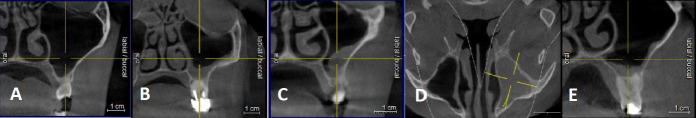
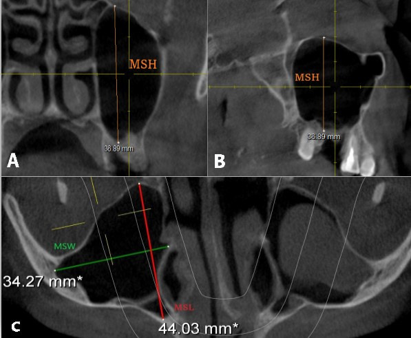
Materials and methods: This retrospective descriptive study analyzed 200 CBCT scans, comprising 400 maxillary sinuses from patients treated at the Faculty of Dentistry University of Sarajevo. The study assessed sinus dimensions, volumes, anatomical features, their relationships with adjacent anatomical structures, and the occurrence of pathological alterations. Furthermore, the patient's gender and dental status were studied in relation to these features. The sinus measurements were performed with Sidexis 4 software (Dentsply Sirona, Germany), which is intended for precise linear measurements in three orthogonal planes.
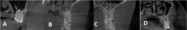
Results: Significant differences were found in sinus width between patients with full dentition and those with partial or complete edentulism (p<0.01). Male participants exhibited larger mean sinus dimensions compared to females in all dimensions. The maxillary sinus floor was inferior to the nasal floor in 91.5% of cases. Various types of sinus membrane abnormalities were observed, with normal membrane thickness in 53.75% of cases. Sinus septa were most frequently located on the roof of the sinus, and their frequency varied significantly among patients with different dentition statuses. The study also identified 274 Haller cells and documented several pathological changes, with mucosal thickening exceeding 3 mm being the most common alteration.
Conclusion: This radiographic study of Bosnian and Herzegovian population revealed significant anatomical variations and pathological changes in maxillary sinuses, thus emphasizing the importance of careful preoperative evaluation using CBCT for surgical planning in the posterior maxillary area. The findings highlight gender-based differences in sinus volumes, the impact of dentition status on sinus anatomy, and the prevalence of various pathological conditions, thus contributing to valuable insights in the field of maxillofacial radiology.
期刊介绍:
The Acta Stomatologica Croatica (ASCRO) is a leading scientific non-profit journal in the field of dental, oral and cranio-facial sciences during the past 44 years in Croatia. ASCRO publishes original scientific and clinical papers, preliminary communications, case reports, book reviews, letters to the editor and news. Review articles are published by invitation from the Editor-in-Chief by acclaimed professionals in distinct fields of dental medicine. All manuscripts are subjected to peer review process.

 求助内容:
求助内容: 应助结果提醒方式:


