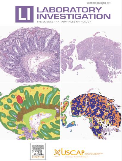
Role of Reserve Cells in Metaplasia and the Development of Human Papillomavirus–Associated High-Grade Squamous Intraepithelial Lesions at the Cervical Transformation Zone
IF 5.1
2区 医学
Q1 MEDICINE, RESEARCH & EXPERIMENTAL
引用次数: 0
Abstract
Squamous cervical cancers generally arise as a result of persistent infection with high-risk human papillomaviruses (hrHPVs) and occur near the squamocolumnar junction (SCJ) and within the transformation zone (TZ). The susceptibility of the TZ to HPV-related carcinogenesis appears linked to epithelial cell plasticity, with squamous metaplasia originating from a specialized stem cell population at this site. Two alternative cell populations have been implicated: keratin (K)7+ve cuboidal cells located at the SCJ vs a more broadly distributed K17+ve cervical reserve cell population. To distinguish between the hypotheses, we utilized multiplex immunofluorescence and large-scale digital imaging to map cell populations at the TZ of 165 women with and without hrHPV infections. Our results did not reveal a distinct population of K7+ cuboidal cells at the SCJ but found instead that the cuboidal and columnar cells of the TZ express K7 and K8 throughout and lack the p63 transcription factor required for epithelial stratification. Squamous metaplasia and reserve cells, which are defined by their subcolumnar location and pattern of biomarker expression (K5/K17/P63), were conspicuous at cervical crypt entrances within the TZ extending proximally toward the endocervix. In HPV-infected tissue, crypt-entrance regions with thin high-grade squamous intraepithelial lesion pathology showed prominent expression of hrHPV E6/E7 mRNA, as detected by fluorescence in situ hybridization, and p16/MCM expression, with infection also apparent in neighboring reserve cells. In some instances, normal/uninfected reserve cells (E6/E7 mRNA−ve) and squamous metaplasia were not only seen close to these regions of hrHPV infection but also extended well beyond the infected area both laterally and by depth. Our results confirm that the reserve cells underneath the columnar epithelia at TZ have the potential to undergo malignant squamous transformation via reserve cell proliferation, in agreement with previous histopathological studies. These translational findings highlight the importance of understanding the molecular biology of the epithelial sites where HPV cancers develop and suggest that in high-risk individuals, treatment strategies should target a wider area than previously thought.
储备细胞在人乳头瘤病毒相关宫颈转化区高级别鳞状上皮内病变化生和发展中的作用
宫颈鳞状癌通常是由于高风险人乳头瘤病毒(hrhpv)持续感染而引起的,发生在鳞状柱交界处(SCJ)附近和转化区(TZ)内。TZ对hpv相关癌变的易感性似乎与上皮细胞的可塑性有关,鳞状化生起源于该部位的特殊干细胞群。两种不同的细胞群涉及:角蛋白(K)7+ve立方体细胞位于SCJ和更广泛分布的K17+ve宫颈储备细胞群。为了区分这些假设,我们利用多重免疫荧光和大规模数字成像来绘制165名感染和未感染hrHPV的妇女TZ的细胞群。我们的研究结果并没有显示SCJ中有明显的K7+立方细胞群,而是发现TZ的立方细胞和柱状细胞在整个细胞中表达K7和K8,并且缺乏上皮分层所需的p63转录因子。鳞状化生和储备细胞由其柱状下位置和生物标志物表达模式(K5/K17/P63)定义,在TZ内近端向宫颈内延伸的宫颈隐窝入口明显。在hpv感染的组织中,具有薄的高级别鳞状上皮内病变病理的隐窝入口区,荧光原位杂交检测到hrHPV E6/E7 mRNA和p16/MCM表达显著,邻近的储备细胞也明显感染。在某些情况下,正常/未感染的储备细胞(E6/E7 mRNA - ve)和鳞状化生不仅在这些hrHPV感染区域附近可见,而且在侧面和深度上都远远超出了感染区域。我们的研究结果证实,TZ柱状上皮下的储备细胞有可能通过储备细胞增殖发生恶性鳞状转化,这与先前的组织病理学研究一致。这些转化研究结果强调了了解HPV癌症发生的上皮部位的分子生物学的重要性,并表明在高危人群中,治疗策略应该针对比以前认为的更广泛的区域。
本文章由计算机程序翻译,如有差异,请以英文原文为准。
求助全文
约1分钟内获得全文
求助全文
来源期刊

Laboratory Investigation
医学-病理学
CiteScore
8.30
自引率
0.00%
发文量
125
审稿时长
2 months
期刊介绍:
Laboratory Investigation is an international journal owned by the United States and Canadian Academy of Pathology. Laboratory Investigation offers prompt publication of high-quality original research in all biomedical disciplines relating to the understanding of human disease and the application of new methods to the diagnosis of disease. Both human and experimental studies are welcome.
 求助内容:
求助内容: 应助结果提醒方式:


