Cone Density Changes After Repeated Low-Level Red Light Treatment in Children With Myopia

IF 7.8 1区 医学 Q1 OPHTHALMOLOGY
Xinyi Liao, Jifeng Yu, Yuzhuo Fan, Yixuan Zhang, Yan Li, Xuewei Li, Hongxin Song, Kai Wang
{"title":"Cone Density Changes After Repeated Low-Level Red Light Treatment in Children With Myopia","authors":"Xinyi Liao, Jifeng Yu, Yuzhuo Fan, Yixuan Zhang, Yan Li, Xuewei Li, Hongxin Song, Kai Wang","doi":"10.1001/jamaophthalmol.2025.0835","DOIUrl":null,"url":null,"abstract":"ImportanceRepeated low-level red light (RLRL) therapy has emerged as a potential intervention for controlling myopia progression in children. However, its long-term effects on retinal photoreceptors remain relatively unknown.ObjectiveTo evaluate changes associated with RLRL therapy on cone photoreceptor density in children with myopia using high-resolution adaptive optics scanning laser ophthalmoscopy (AOSLO).Design, Setting, and ParticipantsThis retrospective multicenter cohort study analyzed data collected from January to March 2024, focusing on Chinese children with myopia. All participants were recruited through questionnaires. Cone density measurements were obtained from AOSLO retinal images. Children with myopia aged 5 to 14 years recruited from the pediatric ophthalmology clinic during routine eye examinations were included in the study and assigned to the RLRL group or the control group. Inclusion criteria were spherical equivalent refraction below −6.00 diopters (D) and best-corrected visual acuity ≥20/20.ExposuresCone density measurement with AOSLO retinal images.Main Outcomes and MeasuresCone photoreceptor density was measured along 4 retinal meridians from central fovea to 4° eccentricity on AOSLO. Fundus abnormalities were assessed using AOSLO, optical coherence tomography (OCT), and fundus photography. Image evaluators were masked to group allocation.ResultsA total of 99 children with myopia were included in this analysis: 52 (97 eyes; mean [SD] age, 10.3 [1.9] years; 27 female [51.9%]) in the RLRL group and 47 (74 eyes; mean [SD] age, 9.8 [2.1] years; 25 male [53.2%]) in the control group. RLRL users showed decreased cone density within 0.5-mm eccentricity from the foveal center, most notably in the temporal region. At 0.3-mm temporal eccentricity, the RLRL group showed a mean difference of −2.1 × 10<jats:sup>3</jats:sup> cells/mm<jats:sup>2</jats:sup> compared with controls (95% CI, −3.68 to −0.59 × 10<jats:sup>3</jats:sup> cells/mm<jats:sup>2</jats:sup>; <jats:italic>P</jats:italic> = .003). A total of 11 eyes exhibited abnormal low-frequency, high-brightness signals near the fovea. The odds ratio of abnormal signals in RLRL users compared with nonusers was 7.23 (95% CI, 1.15-303.45; Fisher exact test, <jats:italic>P</jats:italic> = .02). One participant had relatively small cystoid abnormalities on OCT in the ganglion cell layer, which resolved 3 months after discontinuing RLRL therapy.Conclusions and RelevanceResults of this cohort study suggest that RLRL therapy for at least 1 year was associated with reduced cone density in the paracentral fovea and other subtle retinal abnormalities in some children receiving this therapy for myopia control. These findings support the need for further research to evaluate longer-term safety of RLRL therapy in similar individuals.","PeriodicalId":14518,"journal":{"name":"JAMA ophthalmology","volume":"23 1","pages":""},"PeriodicalIF":7.8000,"publicationDate":"2025-04-24","publicationTypes":"Journal Article","fieldsOfStudy":null,"isOpenAccess":false,"openAccessPdf":"","citationCount":"0","resultStr":null,"platform":"Semanticscholar","paperid":null,"PeriodicalName":"JAMA ophthalmology","FirstCategoryId":"3","ListUrlMain":"https://doi.org/10.1001/jamaophthalmol.2025.0835","RegionNum":1,"RegionCategory":"医学","ArticlePicture":[],"TitleCN":null,"AbstractTextCN":null,"PMCID":null,"EPubDate":"","PubModel":"","JCR":"Q1","JCRName":"OPHTHALMOLOGY","Score":null,"Total":0}
引用次数: 0

Abstract

ImportanceRepeated low-level red light (RLRL) therapy has emerged as a potential intervention for controlling myopia progression in children. However, its long-term effects on retinal photoreceptors remain relatively unknown.ObjectiveTo evaluate changes associated with RLRL therapy on cone photoreceptor density in children with myopia using high-resolution adaptive optics scanning laser ophthalmoscopy (AOSLO).Design, Setting, and ParticipantsThis retrospective multicenter cohort study analyzed data collected from January to March 2024, focusing on Chinese children with myopia. All participants were recruited through questionnaires. Cone density measurements were obtained from AOSLO retinal images. Children with myopia aged 5 to 14 years recruited from the pediatric ophthalmology clinic during routine eye examinations were included in the study and assigned to the RLRL group or the control group. Inclusion criteria were spherical equivalent refraction below −6.00 diopters (D) and best-corrected visual acuity ≥20/20.ExposuresCone density measurement with AOSLO retinal images.Main Outcomes and MeasuresCone photoreceptor density was measured along 4 retinal meridians from central fovea to 4° eccentricity on AOSLO. Fundus abnormalities were assessed using AOSLO, optical coherence tomography (OCT), and fundus photography. Image evaluators were masked to group allocation.ResultsA total of 99 children with myopia were included in this analysis: 52 (97 eyes; mean [SD] age, 10.3 [1.9] years; 27 female [51.9%]) in the RLRL group and 47 (74 eyes; mean [SD] age, 9.8 [2.1] years; 25 male [53.2%]) in the control group. RLRL users showed decreased cone density within 0.5-mm eccentricity from the foveal center, most notably in the temporal region. At 0.3-mm temporal eccentricity, the RLRL group showed a mean difference of −2.1 × 10³ cells/mm2 compared with controls (95% CI, −3.68 to −0.59 × 10³ cells/mm2; P = .003). A total of 11 eyes exhibited abnormal low-frequency, high-brightness signals near the fovea. The odds ratio of abnormal signals in RLRL users compared with nonusers was 7.23 (95% CI, 1.15-303.45; Fisher exact test, P = .02). One participant had relatively small cystoid abnormalities on OCT in the ganglion cell layer, which resolved 3 months after discontinuing RLRL therapy.Conclusions and RelevanceResults of this cohort study suggest that RLRL therapy for at least 1 year was associated with reduced cone density in the paracentral fovea and other subtle retinal abnormalities in some children receiving this therapy for myopia control. These findings support the need for further research to evaluate longer-term safety of RLRL therapy in similar individuals.
近视儿童反复低强度红光治疗后视锥细胞密度的变化
重要性重复低强度红光(RLRL)疗法已成为控制儿童近视发展的一种潜在干预措施。设计、设置和参与者这项回顾性多中心队列研究分析了 2024 年 1 月至 3 月收集的数据,主要针对中国的近视儿童。所有参与者都是通过问卷调查招募的。锥体密度的测量数据来自 AOSLO 视网膜图像。在常规眼科检查中从儿童眼科诊所招募的5至14岁近视儿童被纳入研究,并被分配到RLRL组或对照组。纳入标准为球面等效屈光度低于-6.00屈光度(D),且最佳矫正视力≥20/20。主要结果和测量方法用AOSLO视网膜图像测量从中心眼窝到4°偏心沿4条视网膜经线的锥体光感受器密度。使用 AOSLO、光学相干断层扫描 (OCT) 和眼底摄影评估眼底异常。结果 共有 99 名近视儿童参与了此次分析:RLRL 组 52 名(97 只眼睛;平均 [SD] 年龄,10.3 [1.9] 岁;27 名女性 [51.9%]),对照组 47 名(74 只眼睛;平均 [SD] 年龄,9.8 [2.1] 岁;25 名男性 [53.2%])。RLRL 使用者的视锥密度在距离眼窝中心 0.5 mm 偏心率范围内有所下降,最明显的是在颞区。与对照组相比,RLRL 组在 0.3 毫米颞偏心率处的平均差异为 -2.1 × 10³ cells/mm2 (95% CI, -3.68 to -0.59 × 10³ cells/mm2; P = .003)。共有 11 只眼睛在眼窝附近出现低频、高亮度异常信号。RLRL使用者与非使用者相比,出现异常信号的几率比为7.23(95% CI,1.15-303.45;费雪精确检验,P = .02)。结论与相关性这项队列研究的结果表明,RLRL 治疗至少 1 年与一些接受该疗法控制近视的儿童眼窝旁视锥密度降低及其他细微视网膜异常有关。这些发现表明,有必要开展进一步研究,以评估 RLRL疗法在类似人群中的长期安全性。
本文章由计算机程序翻译,如有差异,请以英文原文为准。
求助全文
约1分钟内获得全文 求助全文
来源期刊
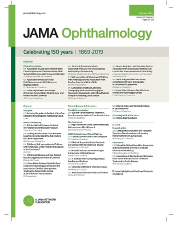
JAMA ophthalmology
JAMA ophthalmology OPHTHALMOLOGY-
CiteScore
13.20
自引率
3.70%
发文量
340
期刊介绍: JAMA Ophthalmology, with a rich history of continuous publication since 1869, stands as a distinguished international, peer-reviewed journal dedicated to ophthalmology and visual science. In 2019, the journal proudly commemorated 150 years of uninterrupted service to the field. As a member of the esteemed JAMA Network, a consortium renowned for its peer-reviewed general medical and specialty publications, JAMA Ophthalmology upholds the highest standards of excellence in disseminating cutting-edge research and insights. Join us in celebrating our legacy and advancing the frontiers of ophthalmology and visual science.
×
引用
GB/T 7714-2015
复制
MLA
复制
APA
复制
导出至
BibTeX EndNote RefMan NoteFirst NoteExpress
×
提示
您的信息不完整,为了账户安全,请先补充。
现在去补充
×
提示
您因"违规操作"
具体请查看互助需知
我知道了
×
提示
确定
请完成安全验证×
copy
已复制链接
快去分享给好友吧!
我知道了
右上角分享
点击右上角分享
0
联系我们:info@booksci.cn Book学术提供免费学术资源搜索服务,方便国内外学者检索中英文文献。致力于提供最便捷和优质的服务体验。 Copyright © 2023 布克学术 All rights reserved.
京ICP备2023020795号-1
ghs 京公网安备 11010802042870号
Book学术文献互助
Book学术文献互助群
群 号:481959085
Book学术官方微信