Khin Yadanar Kyaw, Min Thant Lwin, Alistair Gummow, Antonia Ugur
{"title":"Multi-disciplinary collaboration in diagnosing thymic hyperplasia in a Graves' disease patient.","authors":"Khin Yadanar Kyaw, Min Thant Lwin, Alistair Gummow, Antonia Ugur","doi":"10.1530/EDM-24-0125","DOIUrl":null,"url":null,"abstract":"<p><strong>Summary: </strong>Thymic hyperplasia in Graves' disease is rarely identified due to the absence of routine imaging, but not uncommonly present. It is usually seen when imaging is performed for other reasons. Despite thymic hyperplasia becoming a more commonly identified occurrence, follow-up imaging scans and multi-disciplinary team (MDT) approach are still recommended to distinguish this benign transformation from more significant differentials. These steps can lead to distress in patients. Therefore, clinicians and radiologists being aware of this correlation between thymic hyperplasia and Graves' disease can add reassurances about the most likely diagnosis whilst the patient is undergoing limited further investigation to rule out differentials and subsequently, avoid unnecessary intervention. Here, we report a case of Graves' disease with thymic hyperplasia in a young woman who initially presented with non-specific eye symptoms and incidental mediastinal mass, in which involvement of multiple speciality teams was important to rule out thymoma and myasthenia gravis (MG).</p><p><strong>Learning points: </strong>Although Graves' disease with thymic hyperplasia is not uncommon, it is sometimes difficult to diagnose with one imaging scan due to the overlap of radiological characteristics of other important differentials; an MDT discussion and further imaging scans are needed to confirm the diagnosis in some cases. Getting MDT involvement early would quickly assist in ruling out more significant differentials and avoid unnecessary surgical intervention by concluding thymic hyperplasia. Clinicians having knowledge on the relation between Graves' disease and thymic hyperplasia may reassure the patient by explaining the possible resolution with treatment, while awaiting further MDT discussion. To rule out ocular MG in Graves' disease patients, additional investigations and neurology referral are often required as the serum antibody tests are less sensitive in ocular MG than generalised MG.</p>","PeriodicalId":37467,"journal":{"name":"Endocrinology, Diabetes and Metabolism Case Reports","volume":"2025 2","pages":""},"PeriodicalIF":0.7000,"publicationDate":"2025-04-08","publicationTypes":"Journal Article","fieldsOfStudy":null,"isOpenAccess":false,"openAccessPdf":"https://www.ncbi.nlm.nih.gov/pmc/articles/PMC12272821/pdf/","citationCount":"0","resultStr":null,"platform":"Semanticscholar","paperid":null,"PeriodicalName":"Endocrinology, Diabetes and Metabolism Case Reports","FirstCategoryId":"1085","ListUrlMain":"https://doi.org/10.1530/EDM-24-0125","RegionNum":0,"RegionCategory":null,"ArticlePicture":[],"TitleCN":null,"AbstractTextCN":null,"PMCID":null,"EPubDate":"2025/4/1 0:00:00","PubModel":"Print","JCR":"Q4","JCRName":"ENDOCRINOLOGY & METABOLISM","Score":null,"Total":0}
引用次数: 0
Abstract
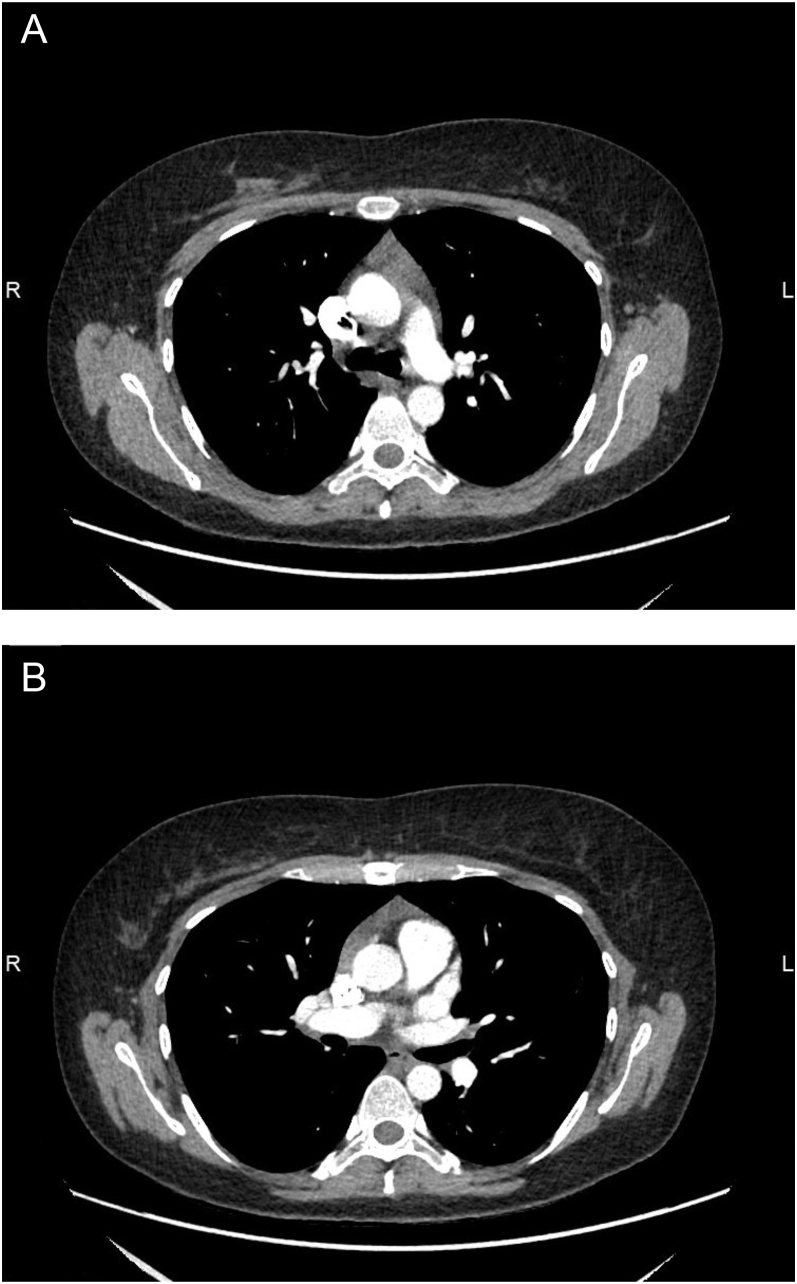
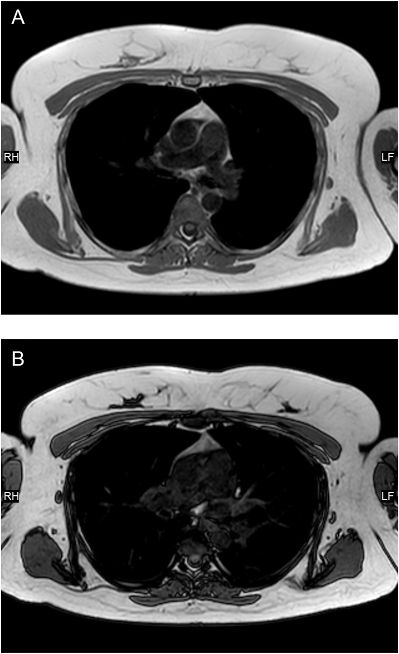
Summary: Thymic hyperplasia in Graves' disease is rarely identified due to the absence of routine imaging, but not uncommonly present. It is usually seen when imaging is performed for other reasons. Despite thymic hyperplasia becoming a more commonly identified occurrence, follow-up imaging scans and multi-disciplinary team (MDT) approach are still recommended to distinguish this benign transformation from more significant differentials. These steps can lead to distress in patients. Therefore, clinicians and radiologists being aware of this correlation between thymic hyperplasia and Graves' disease can add reassurances about the most likely diagnosis whilst the patient is undergoing limited further investigation to rule out differentials and subsequently, avoid unnecessary intervention. Here, we report a case of Graves' disease with thymic hyperplasia in a young woman who initially presented with non-specific eye symptoms and incidental mediastinal mass, in which involvement of multiple speciality teams was important to rule out thymoma and myasthenia gravis (MG).
Learning points: Although Graves' disease with thymic hyperplasia is not uncommon, it is sometimes difficult to diagnose with one imaging scan due to the overlap of radiological characteristics of other important differentials; an MDT discussion and further imaging scans are needed to confirm the diagnosis in some cases. Getting MDT involvement early would quickly assist in ruling out more significant differentials and avoid unnecessary surgical intervention by concluding thymic hyperplasia. Clinicians having knowledge on the relation between Graves' disease and thymic hyperplasia may reassure the patient by explaining the possible resolution with treatment, while awaiting further MDT discussion. To rule out ocular MG in Graves' disease patients, additional investigations and neurology referral are often required as the serum antibody tests are less sensitive in ocular MG than generalised MG.
期刊介绍:
Endocrinology, Diabetes & Metabolism Case Reports publishes case reports on common and rare conditions in all areas of clinical endocrinology, diabetes and metabolism. Articles should include clear learning points which readers can use to inform medical education or clinical practice. The types of cases of interest to Endocrinology, Diabetes & Metabolism Case Reports include: -Insight into disease pathogenesis or mechanism of therapy - Novel diagnostic procedure - Novel treatment - Unique/unexpected symptoms or presentations of a disease - New disease or syndrome: presentations/diagnosis/management - Unusual effects of medical treatment - Error in diagnosis/pitfalls and caveats


 求助内容:
求助内容: 应助结果提醒方式:


