Electroacupuncture Improves Ovarian Function in Rats With Tripterygium Glycoside-Induced Diminished Ovarian Reserve by Promoting the Polarization of M2 Macrophages and Inhibiting Inflammatory Responses.
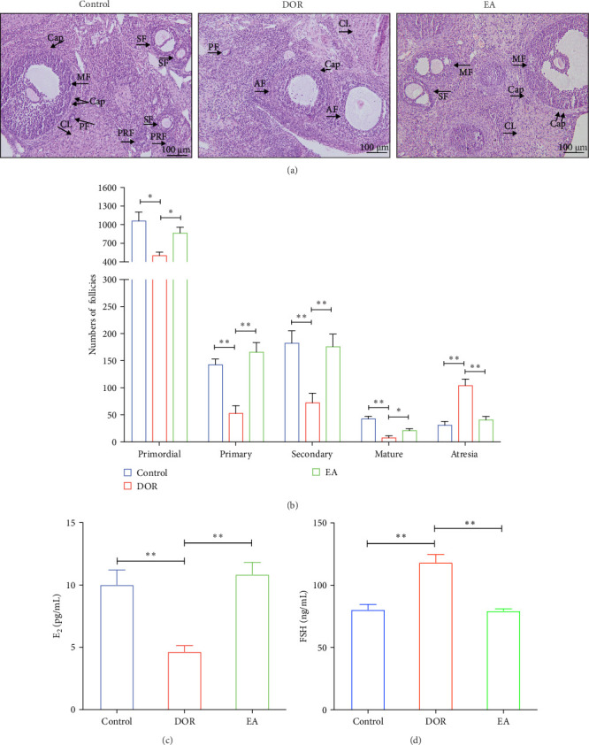
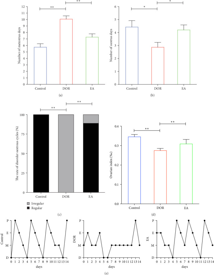
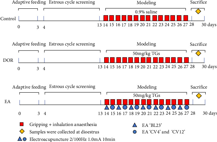
{"title":"Electroacupuncture Improves Ovarian Function in Rats With <i>Tripterygium</i> Glycoside-Induced Diminished Ovarian Reserve by Promoting the Polarization of M2 Macrophages and Inhibiting Inflammatory Responses.","authors":"Jia Luo, Yantong Qin, Yaoyao Zhu, Yaoli Yin, Meihong Shen","doi":"10.1155/mi/1694470","DOIUrl":null,"url":null,"abstract":"<p><p>Immunoinflammatory responses and macrophage polarisation are crucial for maintaining ovarian function. Moreover, electroacupuncture (EA) has been shown to protect ovarian function. However, the mechanisms by which EA improves ovarian function, including its effects on immunoinflammatory responses and macrophage polarisation, have not been determined. This study aimed to investigate the protective effects of EA on ovarian function in rats with diminished ovarian reserve (DOR) and to elucidate the regulatory mechanisms underlying inflammation and M1 and M2 macrophage polarisation. DOR models were established through the intragastric administration of 50 mg/kg <i>Tripterygium</i> glycoside suspension (TGs) for 14 consecutive days. The EA group received treatment at 2/100 Hz and 1.0 mA for 10 min using acupoints BL23, CV4 and CV12 for 14 days. Following the intervention, we employed various methodologies, including haematoxylin-eosin (H&E) staining, enzyme-linked immunosorbent assay (ELISA), flow cytometry, immunohistochemical (IHC) staining, western blotting and quantitative reverse transcriptase-polymerase chain reaction (PCR), to assess ovarian function, inflammatory factors and the expression levels of M1 and M2 macrophage-related factors. EA intervention reduced the oestrous cycle disorder rate in the rats compared with that in the DOR group, leading to an increase in growing follicles, a reduction in atretic follicles (AFs) and an enhancement of both the capillary (Cap) network and corpus luteum (CL) structure. This intervention also resulted in decreased serum levels of follicle-stimulating hormone (FSH), interferon-<i>γ</i> (IFN-<i>γ</i>) and tumour necrosis factor-<i>α</i> (TNF-<i>α</i>), along with increased levels of oestradiol (E<sub>2</sub>), interleukin-4 (IL-4) and interleukin-10 (IL-10). Furthermore, the number of M2 macrophages in the spleen increased, which was accompanied by elevated arginase 1 (Arg1) and decreased inducible nitric oxide synthase (iNOS) expression in the ovarian tissues. In summary, EA can restore the impaired ovarian function caused by TGs by promoting M2 macrophage polarisation and inhibiting inflammatory responses.</p>","PeriodicalId":18371,"journal":{"name":"Mediators of Inflammation","volume":"2025 ","pages":"1694470"},"PeriodicalIF":4.2000,"publicationDate":"2025-03-31","publicationTypes":"Journal Article","fieldsOfStudy":null,"isOpenAccess":false,"openAccessPdf":"https://www.ncbi.nlm.nih.gov/pmc/articles/PMC11976048/pdf/","citationCount":"0","resultStr":null,"platform":"Semanticscholar","paperid":null,"PeriodicalName":"Mediators of Inflammation","FirstCategoryId":"3","ListUrlMain":"https://doi.org/10.1155/mi/1694470","RegionNum":3,"RegionCategory":"医学","ArticlePicture":[],"TitleCN":null,"AbstractTextCN":null,"PMCID":null,"EPubDate":"2025/1/1 0:00:00","PubModel":"eCollection","JCR":"Q2","JCRName":"CELL BIOLOGY","Score":null,"Total":0}
引用次数: 0
Abstract
Immunoinflammatory responses and macrophage polarisation are crucial for maintaining ovarian function. Moreover, electroacupuncture (EA) has been shown to protect ovarian function. However, the mechanisms by which EA improves ovarian function, including its effects on immunoinflammatory responses and macrophage polarisation, have not been determined. This study aimed to investigate the protective effects of EA on ovarian function in rats with diminished ovarian reserve (DOR) and to elucidate the regulatory mechanisms underlying inflammation and M1 and M2 macrophage polarisation. DOR models were established through the intragastric administration of 50 mg/kg Tripterygium glycoside suspension (TGs) for 14 consecutive days. The EA group received treatment at 2/100 Hz and 1.0 mA for 10 min using acupoints BL23, CV4 and CV12 for 14 days. Following the intervention, we employed various methodologies, including haematoxylin-eosin (H&E) staining, enzyme-linked immunosorbent assay (ELISA), flow cytometry, immunohistochemical (IHC) staining, western blotting and quantitative reverse transcriptase-polymerase chain reaction (PCR), to assess ovarian function, inflammatory factors and the expression levels of M1 and M2 macrophage-related factors. EA intervention reduced the oestrous cycle disorder rate in the rats compared with that in the DOR group, leading to an increase in growing follicles, a reduction in atretic follicles (AFs) and an enhancement of both the capillary (Cap) network and corpus luteum (CL) structure. This intervention also resulted in decreased serum levels of follicle-stimulating hormone (FSH), interferon-γ (IFN-γ) and tumour necrosis factor-α (TNF-α), along with increased levels of oestradiol (E2), interleukin-4 (IL-4) and interleukin-10 (IL-10). Furthermore, the number of M2 macrophages in the spleen increased, which was accompanied by elevated arginase 1 (Arg1) and decreased inducible nitric oxide synthase (iNOS) expression in the ovarian tissues. In summary, EA can restore the impaired ovarian function caused by TGs by promoting M2 macrophage polarisation and inhibiting inflammatory responses.
期刊介绍:
Mediators of Inflammation is a peer-reviewed, Open Access journal that publishes original research and review articles on all types of inflammatory mediators, including cytokines, histamine, bradykinin, prostaglandins, leukotrienes, PAF, biological response modifiers and the family of cell adhesion-promoting molecules.
 求助内容:
求助内容: 应助结果提醒方式:


