Evangelia Kaza, Phillip M Devlin, Desmond O'Farrell, Ivan Buzurovic
{"title":"Skin marker for MR-only surface high-dose-rate brachytherapy.","authors":"Evangelia Kaza, Phillip M Devlin, Desmond O'Farrell, Ivan Buzurovic","doi":"10.5114/jcb.2025.148107","DOIUrl":null,"url":null,"abstract":"<p><strong>Purpose: </strong>Recent advances in surface high-dose-rate (HDR) brachytherapy imaging indicate that flap applicators, human skin, and fibromatosis can be visualized using MRI. Complete MR-only surface brachytherapy workflows would require skin marker identification to define clinical target edges. However, CT markers are not detected on MR images, and common MR markers are unsuitable for continuous surface target tracing. In this paper, we proposed an alternative skin marker that was evaluated for MRI and CT detectability and contourability using a brachytherapy treatment planning system (TPS).</p><p><strong>Material and methods: </strong>Commercially obtained silicone rubber tubes of 2 or 3 mm diameter were taped on the hand of an anthropomorphic phantom, a healthy volunteer, and three palmar fascial fibromatosis patients. Subjects were imaged with an optimized 3D pointwise encoding time reduction with radial acquisition (PETRA) sequence, and a volumetric interpolated breath-hold examination (VIBE) sequence with Dixon reconstruction. Additionally, patients underwent standard CT imaging. Obtained images were reviewed for tube conspicuity, and tubes were tracked on axial views using Oncentra Brachy TPS. Independent tube and muscle reference contours were drawn in MIM for quantitative analysis, considering the three orthogonal imaging planes.</p><p><strong>Results and conclusions: </strong>Silicone rubber tubes were detected with positive signal on PETRA, VIBE, and CT images. Among the MR series, Dixon VIBE fat-only showed the highest contrast against muscle tissue and the best separation from human skin, followed by DIXON opposed-phase. 3 mm diameter tubes were tracked better by TPS than 2 mm diameter ones. Considering MR images in the three orthogonal planes in MIM was more helpful for localizing the entire tube than using axial images only in TPS. All obtained contour shapes generally agreed with the known tube positions. Overall, solid silicone rubber tubes of 3 mm diameter represent a suitable skin marker alternative to CT markers for MR-only surface HDR brachytherapy.</p>","PeriodicalId":51305,"journal":{"name":"Journal of Contemporary Brachytherapy","volume":"17 1","pages":"43-53"},"PeriodicalIF":1.1000,"publicationDate":"2025-02-01","publicationTypes":"Journal Article","fieldsOfStudy":null,"isOpenAccess":false,"openAccessPdf":"https://www.ncbi.nlm.nih.gov/pmc/articles/PMC11966218/pdf/","citationCount":"0","resultStr":null,"platform":"Semanticscholar","paperid":null,"PeriodicalName":"Journal of Contemporary Brachytherapy","FirstCategoryId":"3","ListUrlMain":"https://doi.org/10.5114/jcb.2025.148107","RegionNum":4,"RegionCategory":"医学","ArticlePicture":[],"TitleCN":null,"AbstractTextCN":null,"PMCID":null,"EPubDate":"2025/2/27 0:00:00","PubModel":"Epub","JCR":"Q4","JCRName":"ONCOLOGY","Score":null,"Total":0}
引用次数: 0
Abstract
Purpose: Recent advances in surface high-dose-rate (HDR) brachytherapy imaging indicate that flap applicators, human skin, and fibromatosis can be visualized using MRI. Complete MR-only surface brachytherapy workflows would require skin marker identification to define clinical target edges. However, CT markers are not detected on MR images, and common MR markers are unsuitable for continuous surface target tracing. In this paper, we proposed an alternative skin marker that was evaluated for MRI and CT detectability and contourability using a brachytherapy treatment planning system (TPS).
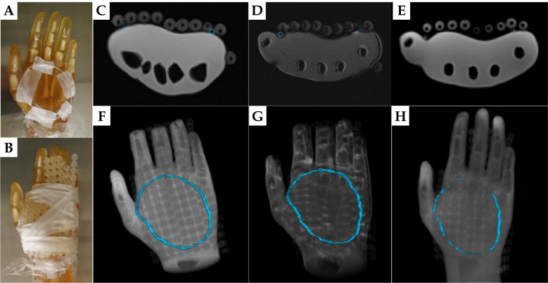
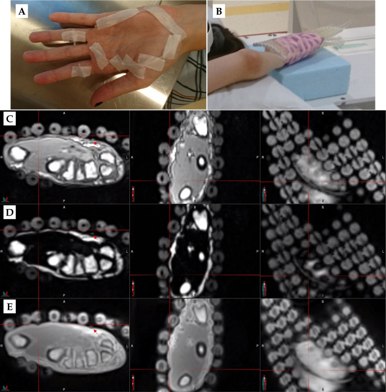
Material and methods: Commercially obtained silicone rubber tubes of 2 or 3 mm diameter were taped on the hand of an anthropomorphic phantom, a healthy volunteer, and three palmar fascial fibromatosis patients. Subjects were imaged with an optimized 3D pointwise encoding time reduction with radial acquisition (PETRA) sequence, and a volumetric interpolated breath-hold examination (VIBE) sequence with Dixon reconstruction. Additionally, patients underwent standard CT imaging. Obtained images were reviewed for tube conspicuity, and tubes were tracked on axial views using Oncentra Brachy TPS. Independent tube and muscle reference contours were drawn in MIM for quantitative analysis, considering the three orthogonal imaging planes.
Results and conclusions: Silicone rubber tubes were detected with positive signal on PETRA, VIBE, and CT images. Among the MR series, Dixon VIBE fat-only showed the highest contrast against muscle tissue and the best separation from human skin, followed by DIXON opposed-phase. 3 mm diameter tubes were tracked better by TPS than 2 mm diameter ones. Considering MR images in the three orthogonal planes in MIM was more helpful for localizing the entire tube than using axial images only in TPS. All obtained contour shapes generally agreed with the known tube positions. Overall, solid silicone rubber tubes of 3 mm diameter represent a suitable skin marker alternative to CT markers for MR-only surface HDR brachytherapy.
期刊介绍:
The “Journal of Contemporary Brachytherapy” is an international and multidisciplinary journal that will publish papers of original research as well as reviews of articles. Main subjects of the journal include: clinical brachytherapy, combined modality treatment, advances in radiobiology, hyperthermia and tumour biology, as well as physical aspects relevant to brachytherapy, particularly in the field of imaging, dosimetry and radiation therapy planning. Original contributions will include experimental studies of combined modality treatment, tumor sensitization and normal tissue protection, molecular radiation biology, and clinical investigations of cancer treatment in brachytherapy. Another field of interest will be the educational part of the journal.

 求助内容:
求助内容: 应助结果提醒方式:


