Fariba Davanian, Iman Adibi, Mahnoosh Tajmirriahi, Maryam Monemian, Zahra Zojaji, Ahmadreza Montazerolghaem, Mohammad Amin Asadinia, Seyed Mojtaba Mirghaderi, Seyed Amin Naji Esfahani, Mohammad Kazemi, Mohammad Reza Iravani, Kian Shahriari, Nesa Sharifi, Sadaf Moharreri, Farnaz Sedighin, Hossein Rabbani
{"title":"Isfahan Artificial Intelligence Event 2023: Lesion Segmentation and Localization in Magnetic Resonance Images of Patients with Multiple Sclerosis.","authors":"Fariba Davanian, Iman Adibi, Mahnoosh Tajmirriahi, Maryam Monemian, Zahra Zojaji, Ahmadreza Montazerolghaem, Mohammad Amin Asadinia, Seyed Mojtaba Mirghaderi, Seyed Amin Naji Esfahani, Mohammad Kazemi, Mohammad Reza Iravani, Kian Shahriari, Nesa Sharifi, Sadaf Moharreri, Farnaz Sedighin, Hossein Rabbani","doi":"10.4103/jmss.jmss_55_24","DOIUrl":null,"url":null,"abstract":"<p><strong>Background: </strong>Multiple sclerosis (MS) is one of the most common reasons of neurological disabilities in young adults. The disease occurs when the immune system attacks the central nervous system and destroys the myelin of nervous cells. This results in appearing several lesions in the magnetic resonance (MR) images of patients. Accurate determination of the amount and the place of lesions can help physicians to determine the severity and progress of the disease.</p><p><strong>Method: </strong>Due to the importance of this issue, this challenge has been dedicated to the segmentation and localization of lesions in MR images of patients with MS. The goal was to segment and localize the lesions in the flair MR images of patients as close as possible to the ground truth masks.</p><p><strong>Results: </strong>Several teams sent us their results for the segmentation and localization of lesions in MR images. Most of the teams preferred to use deep learning methods. The methods varied from a simple U-net structure to more complicated networks.</p><p><strong>Conclusion: </strong>The results show that deep learning methods can be useful for segmentation and localization of lesions in MR images. In this study, we briefly described the dataset and the methods of teams attending the competition.</p>","PeriodicalId":37680,"journal":{"name":"Journal of Medical Signals & Sensors","volume":"15 ","pages":"5"},"PeriodicalIF":1.1000,"publicationDate":"2025-02-28","publicationTypes":"Journal Article","fieldsOfStudy":null,"isOpenAccess":false,"openAccessPdf":"https://www.ncbi.nlm.nih.gov/pmc/articles/PMC11970832/pdf/","citationCount":"0","resultStr":null,"platform":"Semanticscholar","paperid":null,"PeriodicalName":"Journal of Medical Signals & Sensors","FirstCategoryId":"1085","ListUrlMain":"https://doi.org/10.4103/jmss.jmss_55_24","RegionNum":0,"RegionCategory":null,"ArticlePicture":[],"TitleCN":null,"AbstractTextCN":null,"PMCID":null,"EPubDate":"2025/1/1 0:00:00","PubModel":"eCollection","JCR":"Q4","JCRName":"ENGINEERING, BIOMEDICAL","Score":null,"Total":0}
引用次数: 0
Abstract
Background: Multiple sclerosis (MS) is one of the most common reasons of neurological disabilities in young adults. The disease occurs when the immune system attacks the central nervous system and destroys the myelin of nervous cells. This results in appearing several lesions in the magnetic resonance (MR) images of patients. Accurate determination of the amount and the place of lesions can help physicians to determine the severity and progress of the disease.
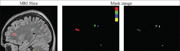
Method: Due to the importance of this issue, this challenge has been dedicated to the segmentation and localization of lesions in MR images of patients with MS. The goal was to segment and localize the lesions in the flair MR images of patients as close as possible to the ground truth masks.
Results: Several teams sent us their results for the segmentation and localization of lesions in MR images. Most of the teams preferred to use deep learning methods. The methods varied from a simple U-net structure to more complicated networks.
Conclusion: The results show that deep learning methods can be useful for segmentation and localization of lesions in MR images. In this study, we briefly described the dataset and the methods of teams attending the competition.
背景:多发性硬化症(MS)是年轻人神经功能障碍的最常见原因之一。当免疫系统攻击中枢神经系统并破坏神经细胞的髓磷脂时,这种疾病就会发生。这导致在患者的磁共振(MR)图像中出现几个病变。准确确定病变的数量和位置可以帮助医生确定疾病的严重程度和进展。方法:由于这个问题的重要性,这个挑战一直致力于ms患者MR图像中病灶的分割和定位,目标是在患者的flair MR图像中尽可能接近ground truth mask的病灶分割和定位。结果:几个团队向我们发送了他们的MR图像中病灶的分割和定位结果。大多数团队更喜欢使用深度学习方法。方法从简单的u型网结构到更复杂的网络都有。结论:深度学习方法可用于MR图像中病灶的分割和定位。在本研究中,我们简要描述了数据集和参赛队伍的方法。
期刊介绍:
JMSS is an interdisciplinary journal that incorporates all aspects of the biomedical engineering including bioelectrics, bioinformatics, medical physics, health technology assessment, etc. Subject areas covered by the journal include: - Bioelectric: Bioinstruments Biosensors Modeling Biomedical signal processing Medical image analysis and processing Medical imaging devices Control of biological systems Neuromuscular systems Cognitive sciences Telemedicine Robotic Medical ultrasonography Bioelectromagnetics Electrophysiology Cell tracking - Bioinformatics and medical informatics: Analysis of biological data Data mining Stochastic modeling Computational genomics Artificial intelligence & fuzzy Applications Medical softwares Bioalgorithms Electronic health - Biophysics and medical physics: Computed tomography Radiation therapy Laser therapy - Education in biomedical engineering - Health technology assessment - Standard in biomedical engineering.

 求助内容:
求助内容: 应助结果提醒方式:


