Cheng-Ting Shih, Ko-Han Lin, Bang-Hung Yang, Chien-Ying Li, Tzu-Lin Lin, Greta S P Mok, Tung-Hsin Wu
{"title":"Deriving tissue physical densities based on Dixon magnetic resonance images and tissue composition prior knowledge for voxel-based internal dosimetry.","authors":"Cheng-Ting Shih, Ko-Han Lin, Bang-Hung Yang, Chien-Ying Li, Tzu-Lin Lin, Greta S P Mok, Tung-Hsin Wu","doi":"10.1186/s40658-025-00737-4","DOIUrl":null,"url":null,"abstract":"<p><strong>Background: </strong>Magnetic resonance (MR) images have been applied in diagnostic and therapeutic nuclear medicine to improve the visualization and characterization of soft tissues and tumors. However, the physical density (ρ) and elemental composition of human tissues required for dosimetric calculation cannot be directly converted from MR images, obstructing MR-based personalized internal dosimetry. In this study, we proposed a method to derive physical densities from Dixon MR images for voxel-based internal dose calculation.</p><p><strong>Methods: </strong>The proposed method defined human tissues as composed of four basic tissues. The physical densities of the human tissues were calculated using the standard tissue composition of the basic tissues and the volume fraction maps calculated from Dixon images. The derived ρ map was applied to calculate the whole-body internal dosimetry using a multiple voxel S-value (MSV) approach. The accuracy of the proposed method in deriving ρ and calculating the internal dose of <sup>18</sup>F-FDG PET imaging was evaluated by comparing with those obtained from computed tomography (CT) images of the same patient and was compared with those obtained using generative adversarial networks (GANs).</p><p><strong>Results: </strong>The proposed method was superior to the GANs in deriving ρ from Dixon MR images and the following internal dose calculation. On average of a validation set, the mean absolute percent errors (MAPEs) of the whole-body ρ derivation and internal dose calculation using the proposed method were 14.28 ± 11.11% and 3.31 ± 0.69%, respectively. The MAPEs were respectively reduced to 5.97 ± 2.51 and 2.75 ± 0.69% after excluding the intestinal gas with different locations in the Dixon MR and CT images.</p><p><strong>Conclusions: </strong>The proposed method could be applied for accurate and efficient personalized internal dosimetry evaluation in MR-integrated nuclear medicine clinical applications.</p>","PeriodicalId":11559,"journal":{"name":"EJNMMI Physics","volume":"12 1","pages":"36"},"PeriodicalIF":3.2000,"publicationDate":"2025-04-07","publicationTypes":"Journal Article","fieldsOfStudy":null,"isOpenAccess":false,"openAccessPdf":"https://www.ncbi.nlm.nih.gov/pmc/articles/PMC11977065/pdf/","citationCount":"0","resultStr":null,"platform":"Semanticscholar","paperid":null,"PeriodicalName":"EJNMMI Physics","FirstCategoryId":"3","ListUrlMain":"https://doi.org/10.1186/s40658-025-00737-4","RegionNum":2,"RegionCategory":"医学","ArticlePicture":[],"TitleCN":null,"AbstractTextCN":null,"PMCID":null,"EPubDate":"","PubModel":"","JCR":"Q2","JCRName":"RADIOLOGY, NUCLEAR MEDICINE & MEDICAL IMAGING","Score":null,"Total":0}
引用次数: 0
Abstract
Background: Magnetic resonance (MR) images have been applied in diagnostic and therapeutic nuclear medicine to improve the visualization and characterization of soft tissues and tumors. However, the physical density (ρ) and elemental composition of human tissues required for dosimetric calculation cannot be directly converted from MR images, obstructing MR-based personalized internal dosimetry. In this study, we proposed a method to derive physical densities from Dixon MR images for voxel-based internal dose calculation.
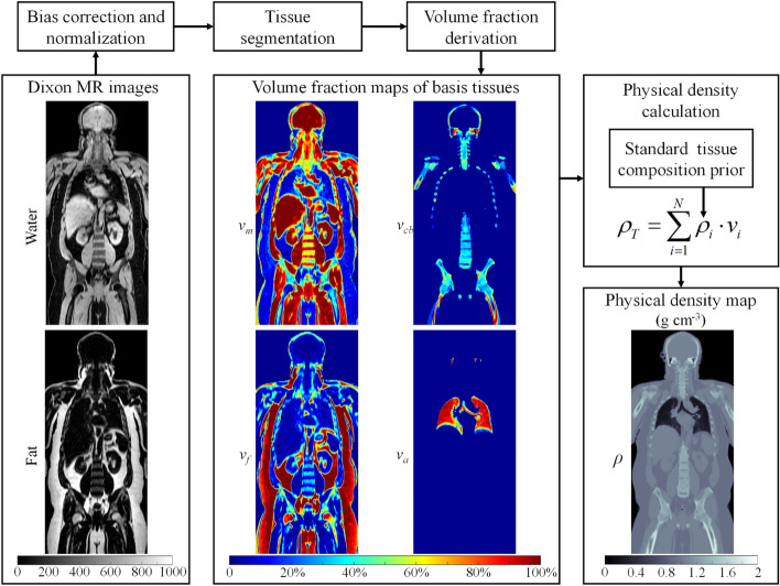
Methods: The proposed method defined human tissues as composed of four basic tissues. The physical densities of the human tissues were calculated using the standard tissue composition of the basic tissues and the volume fraction maps calculated from Dixon images. The derived ρ map was applied to calculate the whole-body internal dosimetry using a multiple voxel S-value (MSV) approach. The accuracy of the proposed method in deriving ρ and calculating the internal dose of 18F-FDG PET imaging was evaluated by comparing with those obtained from computed tomography (CT) images of the same patient and was compared with those obtained using generative adversarial networks (GANs).
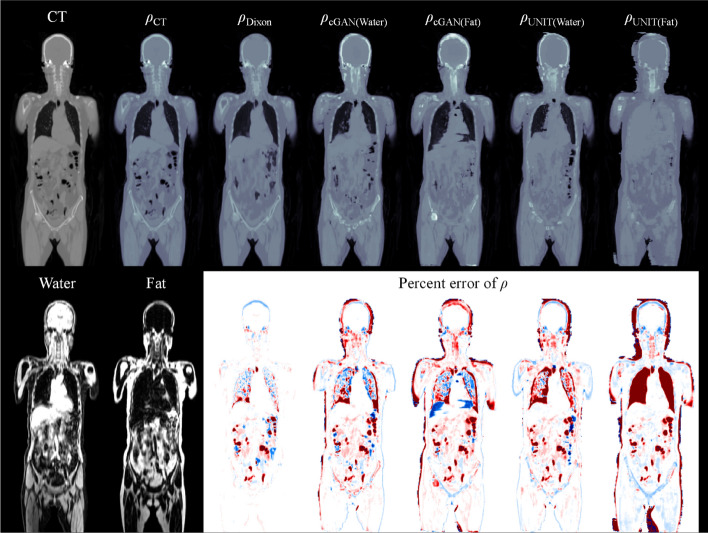
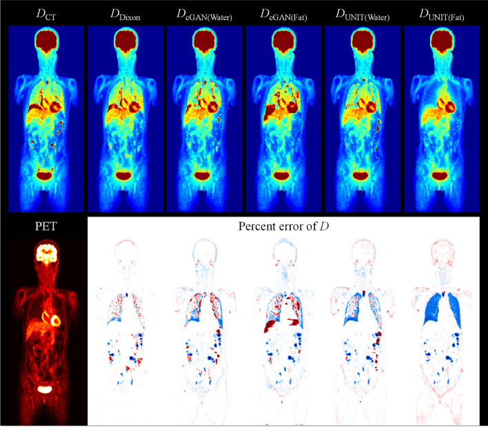
Results: The proposed method was superior to the GANs in deriving ρ from Dixon MR images and the following internal dose calculation. On average of a validation set, the mean absolute percent errors (MAPEs) of the whole-body ρ derivation and internal dose calculation using the proposed method were 14.28 ± 11.11% and 3.31 ± 0.69%, respectively. The MAPEs were respectively reduced to 5.97 ± 2.51 and 2.75 ± 0.69% after excluding the intestinal gas with different locations in the Dixon MR and CT images.
Conclusions: The proposed method could be applied for accurate and efficient personalized internal dosimetry evaluation in MR-integrated nuclear medicine clinical applications.
期刊介绍:
EJNMMI Physics is an international platform for scientists, users and adopters of nuclear medicine with a particular interest in physics matters. As a companion journal to the European Journal of Nuclear Medicine and Molecular Imaging, this journal has a multi-disciplinary approach and welcomes original materials and studies with a focus on applied physics and mathematics as well as imaging systems engineering and prototyping in nuclear medicine. This includes physics-driven approaches or algorithms supported by physics that foster early clinical adoption of nuclear medicine imaging and therapy.

 求助内容:
求助内容: 应助结果提醒方式:


