Ellouise C Adams, Katherine Antel, Jenna L Bailey, Karryn L Brown, Dharshnee R Chetty, David Richardson, Estelle Verburgh
{"title":"Diagnostic use of abdominal ultrasound in detecting extrapulmonary tuberculosis or lymphoma in an HIV-endemic region.","authors":"Ellouise C Adams, Katherine Antel, Jenna L Bailey, Karryn L Brown, Dharshnee R Chetty, David Richardson, Estelle Verburgh","doi":"10.4102/sajhivmed.v26i1.1679","DOIUrl":null,"url":null,"abstract":"<p><strong>Background: </strong>Extrapulmonary tuberculosis (EPTB) is common among people living with HIV (PLWH). Abdominal ultrasound is an accessible investigation, frequently employed to support the diagnosis of EPTB, but may lead to misdiagnoses of diseases with overlapping clinical features, such as lymphoma.</p><p><strong>Objectives: </strong>To describe the abdominal ultrasound features and confirmed diagnoses of patients referred to a biopsy clinic with unexplained lymphadenopathy.</p><p><strong>Method: </strong>This was a retrospective descriptive study of patients attending the peripheral lymph node biopsy clinic at Groote Schuur Hospital between 2017 and 2020, who had abdominal ultrasound examinations while being investigated for unexplained lymphadenopathy. Ultrasound features were compared to the final diagnosis made on the lymph node biopsy.</p><p><strong>Results: </strong>Thirty-four patients were included, most of whom were PLWH (59%). Approximately one-third had a confirmed diagnosis of lymphoma (29%) and approximately one-third had a confirmed diagnosis of tuberculosis (32%). Splenic hypoechoic lesions were more common in patients with lymphoma (64%) than in patients with tuberculosis (46%) and malignancy (17%). Ascites was equally distributed between patients with tuberculosis (36%) and lymphoma (36%). The ultrasound report and confirmed diagnoses agreed in 40% of patients with tuberculosis. Additionally, 36% of patients with confirmed lymphoma were suspected to have tuberculosis based on the abdominal ultrasound.</p><p><strong>Conclusion: </strong>Abdominal ultrasound abnormalities such as splenic hypoechoic lesions, lymphadenopathy, and ascites/pleural effusion have a differential diagnosis including both tuberculosis and lymphoma, and should be investigated accordingly.</p>","PeriodicalId":94212,"journal":{"name":"Southern African journal of HIV medicine","volume":"26 1","pages":"1679"},"PeriodicalIF":2.3000,"publicationDate":"2025-03-21","publicationTypes":"Journal Article","fieldsOfStudy":null,"isOpenAccess":false,"openAccessPdf":"https://www.ncbi.nlm.nih.gov/pmc/articles/PMC11966698/pdf/","citationCount":"0","resultStr":null,"platform":"Semanticscholar","paperid":null,"PeriodicalName":"Southern African journal of HIV medicine","FirstCategoryId":"1085","ListUrlMain":"https://doi.org/10.4102/sajhivmed.v26i1.1679","RegionNum":0,"RegionCategory":null,"ArticlePicture":[],"TitleCN":null,"AbstractTextCN":null,"PMCID":null,"EPubDate":"2025/1/1 0:00:00","PubModel":"eCollection","JCR":"","JCRName":"","Score":null,"Total":0}
引用次数: 0
Abstract
Background: Extrapulmonary tuberculosis (EPTB) is common among people living with HIV (PLWH). Abdominal ultrasound is an accessible investigation, frequently employed to support the diagnosis of EPTB, but may lead to misdiagnoses of diseases with overlapping clinical features, such as lymphoma.
Objectives: To describe the abdominal ultrasound features and confirmed diagnoses of patients referred to a biopsy clinic with unexplained lymphadenopathy.
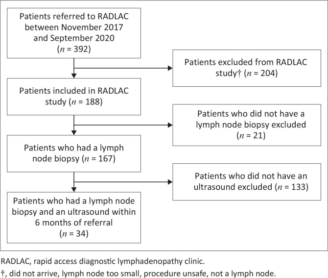
Method: This was a retrospective descriptive study of patients attending the peripheral lymph node biopsy clinic at Groote Schuur Hospital between 2017 and 2020, who had abdominal ultrasound examinations while being investigated for unexplained lymphadenopathy. Ultrasound features were compared to the final diagnosis made on the lymph node biopsy.
Results: Thirty-four patients were included, most of whom were PLWH (59%). Approximately one-third had a confirmed diagnosis of lymphoma (29%) and approximately one-third had a confirmed diagnosis of tuberculosis (32%). Splenic hypoechoic lesions were more common in patients with lymphoma (64%) than in patients with tuberculosis (46%) and malignancy (17%). Ascites was equally distributed between patients with tuberculosis (36%) and lymphoma (36%). The ultrasound report and confirmed diagnoses agreed in 40% of patients with tuberculosis. Additionally, 36% of patients with confirmed lymphoma were suspected to have tuberculosis based on the abdominal ultrasound.
Conclusion: Abdominal ultrasound abnormalities such as splenic hypoechoic lesions, lymphadenopathy, and ascites/pleural effusion have a differential diagnosis including both tuberculosis and lymphoma, and should be investigated accordingly.

 求助内容:
求助内容: 应助结果提醒方式:


