Adam Golda, Beata Bialkowska-Niepon, Tadeusz Zebik
{"title":"Lipomatous Hypertrophy of the Interatrial Septum.","authors":"Adam Golda, Beata Bialkowska-Niepon, Tadeusz Zebik","doi":"10.14740/jmc5108","DOIUrl":null,"url":null,"abstract":"<p><p>The abnormal accumulation of lipid-rich adipose tissue within the interatrial septum (IAS) is the hallmark of lipomatous hypertrophy of the interatrial septum (LHIS), a relatively rare medical condition. To accurately distinguish LHIS, it is essential to recognize the characteristic \"dumbbell\" shape of IAS. Here, we present a case of a 59-year-old woman who was suspected of having cardiac myxoma and was subsequently admitted to our hospital. Transthoracic echocardiography of the patient showed that the IAS had a lack of thickening in the region of the foramen ovale and a hyperechogenic structure in the basal and vault portions of IAS. An abnormal mass located in the IAS anterior to the foramen ovale and not infiltrating the foramen ovale was discovered by computed tomography (CT) scan of the heart. The cardiac magnetic resonance imaging (MRI) confirmed the presence of significant fat deposition within the IAS with sparing of the fossa ovalis, which was consistent with the initial findings. The patient was discharged home with the recommendation of regular visits to the cardiology outpatient clinic for LHIS monitoring. The article presents the visualization of LHIS in consecutive diagnostic modalities, summarizes the actual knowledge of LHIS, and enables proper LHIS diagnosis in patients based on available imaging methods.</p>","PeriodicalId":101328,"journal":{"name":"Journal of medical cases","volume":"16 3","pages":"120-126"},"PeriodicalIF":0.9000,"publicationDate":"2025-03-01","publicationTypes":"Journal Article","fieldsOfStudy":null,"isOpenAccess":false,"openAccessPdf":"https://www.ncbi.nlm.nih.gov/pmc/articles/PMC11954602/pdf/","citationCount":"0","resultStr":null,"platform":"Semanticscholar","paperid":null,"PeriodicalName":"Journal of medical cases","FirstCategoryId":"1085","ListUrlMain":"https://doi.org/10.14740/jmc5108","RegionNum":0,"RegionCategory":null,"ArticlePicture":[],"TitleCN":null,"AbstractTextCN":null,"PMCID":null,"EPubDate":"2025/3/25 0:00:00","PubModel":"Epub","JCR":"","JCRName":"","Score":null,"Total":0}
引用次数: 0
Abstract
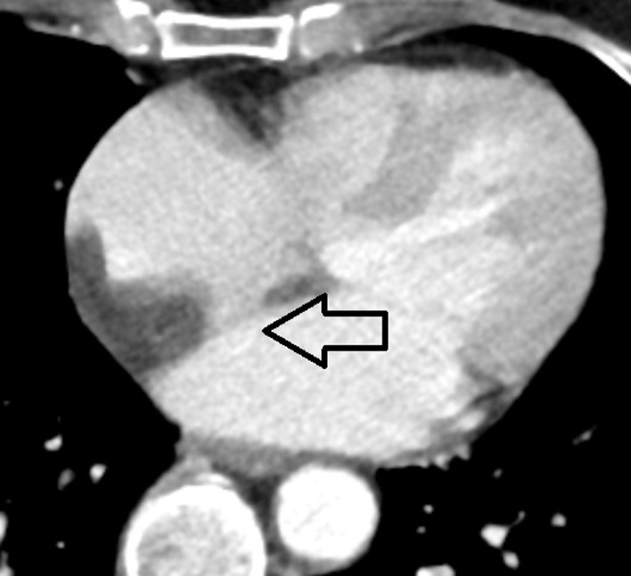
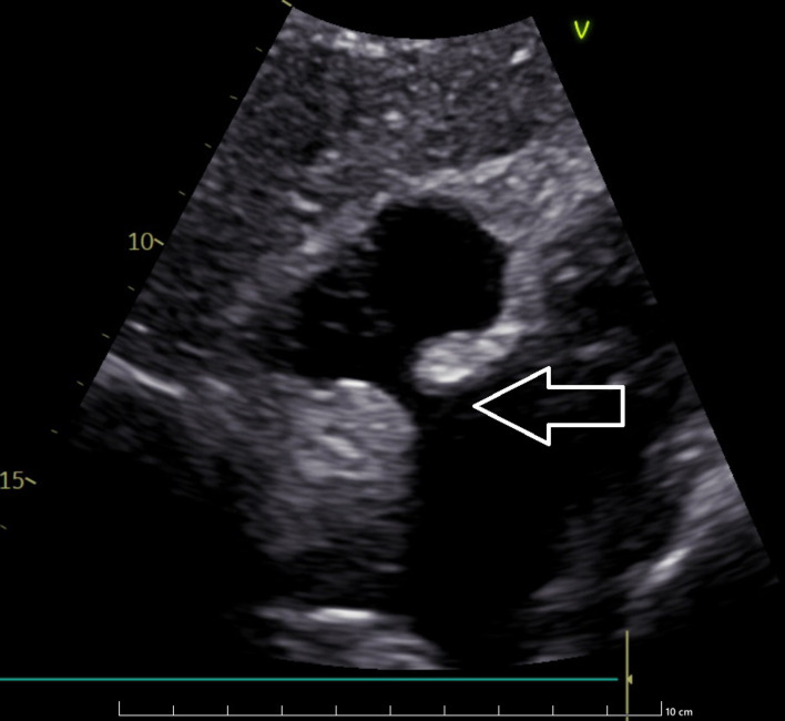
The abnormal accumulation of lipid-rich adipose tissue within the interatrial septum (IAS) is the hallmark of lipomatous hypertrophy of the interatrial septum (LHIS), a relatively rare medical condition. To accurately distinguish LHIS, it is essential to recognize the characteristic "dumbbell" shape of IAS. Here, we present a case of a 59-year-old woman who was suspected of having cardiac myxoma and was subsequently admitted to our hospital. Transthoracic echocardiography of the patient showed that the IAS had a lack of thickening in the region of the foramen ovale and a hyperechogenic structure in the basal and vault portions of IAS. An abnormal mass located in the IAS anterior to the foramen ovale and not infiltrating the foramen ovale was discovered by computed tomography (CT) scan of the heart. The cardiac magnetic resonance imaging (MRI) confirmed the presence of significant fat deposition within the IAS with sparing of the fossa ovalis, which was consistent with the initial findings. The patient was discharged home with the recommendation of regular visits to the cardiology outpatient clinic for LHIS monitoring. The article presents the visualization of LHIS in consecutive diagnostic modalities, summarizes the actual knowledge of LHIS, and enables proper LHIS diagnosis in patients based on available imaging methods.

 求助内容:
求助内容: 应助结果提醒方式:


