Robert H Bardsley, Jasmine Kimber, Kassie McCullagh
{"title":"Primary spinal meningeal melanoma with intramedullary and intradural extramedullary components-a case report.","authors":"Robert H Bardsley, Jasmine Kimber, Kassie McCullagh","doi":"10.1093/bjrcr/uaaf020","DOIUrl":null,"url":null,"abstract":"<p><p>Primary melanomas of the spinal meninges are exceedingly rare. While both intramedullary and extramedullary spinal melanomas have been reported, to the best of our knowledge, this is the first noted case of primary spinal melanoma that has both intramedullary and intradural extramedullary components. We present a case of a 61-year-old female presenting with a 1-year history of lower back pain, bilateral lower extremity pain, and perceived weakness of left foot. Magnetic resonance imaging of the thoracic spine suggested intramedullary and intradural extramedullary mass at levels T8-T12. A T7-T12 laminectomy with resection of the spinal cord mass revealed a pathological diagnosis of primary meningeal melanoma. This case highlights the complexity of diagnosing spinal melanomas, which often mimic more common spinal tumours such as ependymomas, astrocytomas, metastasis, or lymphoma. Often meningeal melanomas require extensive imaging and clinical evaluation to exclude other sites of potential primary melanoma. This case adds to the sparse literature by documenting a rare manifestation and could provide valuable insights into the diagnosis and management of similar cases.</p>","PeriodicalId":45216,"journal":{"name":"BJR Case Reports","volume":"11 2","pages":"uaaf020"},"PeriodicalIF":0.5000,"publicationDate":"2025-03-25","publicationTypes":"Journal Article","fieldsOfStudy":null,"isOpenAccess":false,"openAccessPdf":"https://www.ncbi.nlm.nih.gov/pmc/articles/PMC11954552/pdf/","citationCount":"0","resultStr":null,"platform":"Semanticscholar","paperid":null,"PeriodicalName":"BJR Case Reports","FirstCategoryId":"1085","ListUrlMain":"https://doi.org/10.1093/bjrcr/uaaf020","RegionNum":0,"RegionCategory":null,"ArticlePicture":[],"TitleCN":null,"AbstractTextCN":null,"PMCID":null,"EPubDate":"2025/3/1 0:00:00","PubModel":"eCollection","JCR":"Q4","JCRName":"RADIOLOGY, NUCLEAR MEDICINE & MEDICAL IMAGING","Score":null,"Total":0}
引用次数: 0
Abstract
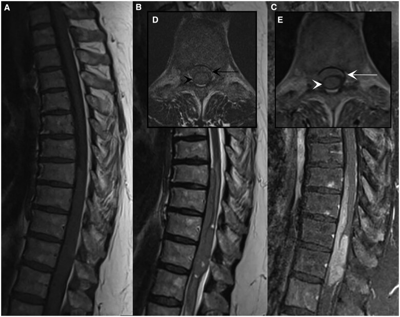
Primary melanomas of the spinal meninges are exceedingly rare. While both intramedullary and extramedullary spinal melanomas have been reported, to the best of our knowledge, this is the first noted case of primary spinal melanoma that has both intramedullary and intradural extramedullary components. We present a case of a 61-year-old female presenting with a 1-year history of lower back pain, bilateral lower extremity pain, and perceived weakness of left foot. Magnetic resonance imaging of the thoracic spine suggested intramedullary and intradural extramedullary mass at levels T8-T12. A T7-T12 laminectomy with resection of the spinal cord mass revealed a pathological diagnosis of primary meningeal melanoma. This case highlights the complexity of diagnosing spinal melanomas, which often mimic more common spinal tumours such as ependymomas, astrocytomas, metastasis, or lymphoma. Often meningeal melanomas require extensive imaging and clinical evaluation to exclude other sites of potential primary melanoma. This case adds to the sparse literature by documenting a rare manifestation and could provide valuable insights into the diagnosis and management of similar cases.

 求助内容:
求助内容: 应助结果提醒方式:


