{"title":"AI-Based Identification Method for Cervical Transformation Zone Within Digital Colposcopy: Development and Multicenter Validation Study.","authors":"Tong Wu, Yuting Wang, Xiaoli Cui, Peng Xue, Youlin Qiao","doi":"10.2196/69672","DOIUrl":null,"url":null,"abstract":"<p><strong>Background: </strong>In low- and middle-income countries, cervical cancer remains a leading cause of death and morbidity for women. Early detection and treatment of precancerous lesions are critical in cervical cancer prevention, and colposcopy is a primary diagnostic tool for identifying cervical lesions and guiding biopsies. The transformation zone (TZ) is where a stratified squamous epithelium develops from the metaplasia of simple columnar epithelium and is the most common site of precancerous lesions. However, inexperienced colposcopists may find it challenging to accurately identify the type and location of the TZ during a colposcopy examination.</p><p><strong>Objective: </strong>This study aims to present an artificial intelligence (AI) method for identifying the TZ to enhance colposcopy examination and evaluate its potential clinical application.</p><p><strong>Methods: </strong>The study retrospectively collected data from 3616 women who underwent colposcopy at 6 tertiary hospitals in China between 2019 and 2021. A dataset from 4 hospitals was collected for model conduction. An independent dataset was collected from the other 2 geographic hospitals to validate model performance. There is no overlap between the training and validation datasets. Anonymized digital records, including each colposcopy image, baseline clinical characteristics, colposcopic findings, and pathological outcomes, were collected. The classification model was proposed as a lightweight neural network with multiscale feature enhancement capabilities and designed to classify the 3 types of TZ. The pretrained FastSAM model was first implemented to identify the location of the new squamocolumnar junction for segmenting the TZ. Overall accuracy, average precision, and recall were evaluated for the classification and segmentation models. The classification performance on the external validation was assessed by sensitivity and specificity.</p><p><strong>Results: </strong>The optimal TZ classification model performed with 83.97% classification accuracy on the test set, which achieved average precision of 91.84%, 89.06%, and 95.62% for types 1, 2, and 3, respectively. The recall and mean average precision of the TZ segmentation model were 0.78 and 0.75, respectively. The proposed model demonstrated outstanding performance in predicting 3 types of the TZ, achieving the sensitivity with 95% CIs for TZ1, TZ2, and TZ3 of 0.78 (0.74-0.81), 0.81 (0.78-0.82), and 0.8 (0.74-0.87), respectively, with specificity with 95% CIs of 0.94 (0.92-0.96), 0.83 (0.81-0.86), and 0.91 (0.89-0.92), based on a comprehensive external dataset of 1335 cases from 2 of the 6 hospitals.</p><p><strong>Conclusions: </strong>Our proposed AI-based identification system classified the type of cervical TZs and delineated their location on multicenter, colposcopic, high-resolution images. The findings of this study have shown its potential to predict TZ types and specific regions accurately. It was developed as a valuable assistant to encourage precise colposcopic examination in clinical practice.</p>","PeriodicalId":45538,"journal":{"name":"JMIR Cancer","volume":"11 ","pages":"e69672"},"PeriodicalIF":2.7000,"publicationDate":"2025-03-31","publicationTypes":"Journal Article","fieldsOfStudy":null,"isOpenAccess":false,"openAccessPdf":"https://www.ncbi.nlm.nih.gov/pmc/articles/PMC11997526/pdf/","citationCount":"0","resultStr":null,"platform":"Semanticscholar","paperid":null,"PeriodicalName":"JMIR Cancer","FirstCategoryId":"1085","ListUrlMain":"https://doi.org/10.2196/69672","RegionNum":0,"RegionCategory":null,"ArticlePicture":[],"TitleCN":null,"AbstractTextCN":null,"PMCID":null,"EPubDate":"","PubModel":"","JCR":"Q2","JCRName":"ONCOLOGY","Score":null,"Total":0}
引用次数: 0
Abstract
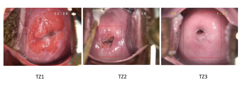
Background: In low- and middle-income countries, cervical cancer remains a leading cause of death and morbidity for women. Early detection and treatment of precancerous lesions are critical in cervical cancer prevention, and colposcopy is a primary diagnostic tool for identifying cervical lesions and guiding biopsies. The transformation zone (TZ) is where a stratified squamous epithelium develops from the metaplasia of simple columnar epithelium and is the most common site of precancerous lesions. However, inexperienced colposcopists may find it challenging to accurately identify the type and location of the TZ during a colposcopy examination.
Objective: This study aims to present an artificial intelligence (AI) method for identifying the TZ to enhance colposcopy examination and evaluate its potential clinical application.
Methods: The study retrospectively collected data from 3616 women who underwent colposcopy at 6 tertiary hospitals in China between 2019 and 2021. A dataset from 4 hospitals was collected for model conduction. An independent dataset was collected from the other 2 geographic hospitals to validate model performance. There is no overlap between the training and validation datasets. Anonymized digital records, including each colposcopy image, baseline clinical characteristics, colposcopic findings, and pathological outcomes, were collected. The classification model was proposed as a lightweight neural network with multiscale feature enhancement capabilities and designed to classify the 3 types of TZ. The pretrained FastSAM model was first implemented to identify the location of the new squamocolumnar junction for segmenting the TZ. Overall accuracy, average precision, and recall were evaluated for the classification and segmentation models. The classification performance on the external validation was assessed by sensitivity and specificity.
Results: The optimal TZ classification model performed with 83.97% classification accuracy on the test set, which achieved average precision of 91.84%, 89.06%, and 95.62% for types 1, 2, and 3, respectively. The recall and mean average precision of the TZ segmentation model were 0.78 and 0.75, respectively. The proposed model demonstrated outstanding performance in predicting 3 types of the TZ, achieving the sensitivity with 95% CIs for TZ1, TZ2, and TZ3 of 0.78 (0.74-0.81), 0.81 (0.78-0.82), and 0.8 (0.74-0.87), respectively, with specificity with 95% CIs of 0.94 (0.92-0.96), 0.83 (0.81-0.86), and 0.91 (0.89-0.92), based on a comprehensive external dataset of 1335 cases from 2 of the 6 hospitals.
Conclusions: Our proposed AI-based identification system classified the type of cervical TZs and delineated their location on multicenter, colposcopic, high-resolution images. The findings of this study have shown its potential to predict TZ types and specific regions accurately. It was developed as a valuable assistant to encourage precise colposcopic examination in clinical practice.


 求助内容:
求助内容: 应助结果提醒方式:


