C J de Wijs, J R Behr, L W J M Streng, M E van der Graaf, F A Harms, E G Mik
{"title":"Automated mitochondrial oxygen consumption (mitoVO<sub>2</sub>) analysis via a bi-directional long short-term memory neural network.","authors":"C J de Wijs, J R Behr, L W J M Streng, M E van der Graaf, F A Harms, E G Mik","doi":"10.1007/s10877-025-01291-1","DOIUrl":null,"url":null,"abstract":"<p><p>Monitoring in vivo mitochondrial oxygen tension (mitoPO<sub>2</sub>) enables the measurement of mitochondrial oxygen consumption (mitoVO<sub>2</sub>), providing deeper insights into the skin's mitochondrial environment. However, current mitoVO<sub>2</sub> analysis often relies on manual identification of start and end points, which introduces substantial inter-user variability. Addressing this limitation is crucial for broader adoption, comparability, and reproducibility across research groups. Therefore, the aim of this study was to develop a neural network-based software that automatically analyzes mitoVO<sub>2</sub>. A Bi-directional Long Short-Term Memory neural network was trained on 125 mitoPO<sub>2</sub> measurement sequences and optimized through Bayesian optimization. It identifies start points and measurement periods, then applies a modified Michaelis-Menten fit to calculate mitoVO<sub>2</sub>. This framework, embedded in automated software, was validated against the consensus of 3 raters. Bayesian optimization yielded an overall network performance of 94.2% on the test set. The neural network identified 91% of mitoVO<sub>2</sub> start points within a ± 5-sample range of the manual consensus. Mean mitoVO<sub>2</sub> values for the consensus and software were 6.56 and 6.63 mmHg s<sup>- 1</sup>, respectively, corresponding to a bias of -0.057 mmHg s<sup>- 1</sup>. Multiple runs of the network on the same dataset produced identical results, confirming consistency and eliminating inter-user variability. The developed neural network-based software automatically and consistently analyzes mitoVO<sub>2</sub> measurements, substantially reducing reliance on subjective judgments. By enabling a standardized approach to mitoVO<sub>2</sub> analysis, this tool improves data comparability and reproducibility across research settings. Future work will focus on further refining precision and extending functionality through multi-center collaborations.</p>","PeriodicalId":15513,"journal":{"name":"Journal of Clinical Monitoring and Computing","volume":" ","pages":"947-956"},"PeriodicalIF":2.2000,"publicationDate":"2025-10-01","publicationTypes":"Journal Article","fieldsOfStudy":null,"isOpenAccess":false,"openAccessPdf":"https://www.ncbi.nlm.nih.gov/pmc/articles/PMC12474646/pdf/","citationCount":"0","resultStr":null,"platform":"Semanticscholar","paperid":null,"PeriodicalName":"Journal of Clinical Monitoring and Computing","FirstCategoryId":"3","ListUrlMain":"https://doi.org/10.1007/s10877-025-01291-1","RegionNum":3,"RegionCategory":"医学","ArticlePicture":[],"TitleCN":null,"AbstractTextCN":null,"PMCID":null,"EPubDate":"2025/3/30 0:00:00","PubModel":"Epub","JCR":"Q2","JCRName":"ANESTHESIOLOGY","Score":null,"Total":0}
引用次数: 0
Abstract
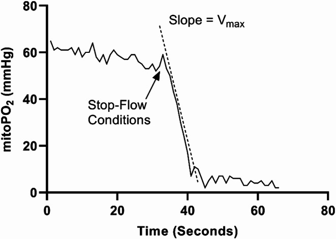
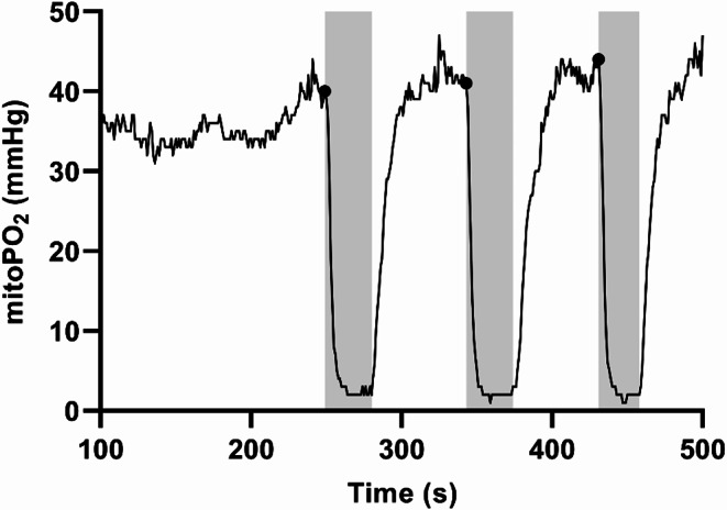
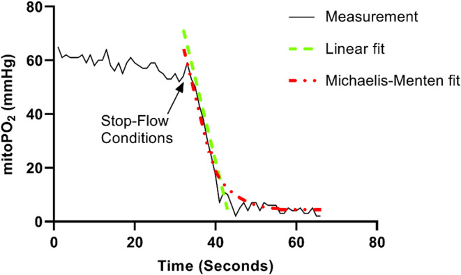
Monitoring in vivo mitochondrial oxygen tension (mitoPO2) enables the measurement of mitochondrial oxygen consumption (mitoVO2), providing deeper insights into the skin's mitochondrial environment. However, current mitoVO2 analysis often relies on manual identification of start and end points, which introduces substantial inter-user variability. Addressing this limitation is crucial for broader adoption, comparability, and reproducibility across research groups. Therefore, the aim of this study was to develop a neural network-based software that automatically analyzes mitoVO2. A Bi-directional Long Short-Term Memory neural network was trained on 125 mitoPO2 measurement sequences and optimized through Bayesian optimization. It identifies start points and measurement periods, then applies a modified Michaelis-Menten fit to calculate mitoVO2. This framework, embedded in automated software, was validated against the consensus of 3 raters. Bayesian optimization yielded an overall network performance of 94.2% on the test set. The neural network identified 91% of mitoVO2 start points within a ± 5-sample range of the manual consensus. Mean mitoVO2 values for the consensus and software were 6.56 and 6.63 mmHg s- 1, respectively, corresponding to a bias of -0.057 mmHg s- 1. Multiple runs of the network on the same dataset produced identical results, confirming consistency and eliminating inter-user variability. The developed neural network-based software automatically and consistently analyzes mitoVO2 measurements, substantially reducing reliance on subjective judgments. By enabling a standardized approach to mitoVO2 analysis, this tool improves data comparability and reproducibility across research settings. Future work will focus on further refining precision and extending functionality through multi-center collaborations.
期刊介绍:
The Journal of Clinical Monitoring and Computing is a clinical journal publishing papers related to technology in the fields of anaesthesia, intensive care medicine, emergency medicine, and peri-operative medicine.
The journal has links with numerous specialist societies, including editorial board representatives from the European Society for Computing and Technology in Anaesthesia and Intensive Care (ESCTAIC), the Society for Technology in Anesthesia (STA), the Society for Complex Acute Illness (SCAI) and the NAVAt (NAVigating towards your Anaestheisa Targets) group.
The journal publishes original papers, narrative and systematic reviews, technological notes, letters to the editor, editorial or commentary papers, and policy statements or guidelines from national or international societies. The journal encourages debate on published papers and technology, including letters commenting on previous publications or technological concerns. The journal occasionally publishes special issues with technological or clinical themes, or reports and abstracts from scientificmeetings. Special issues proposals should be sent to the Editor-in-Chief. Specific details of types of papers, and the clinical and technological content of papers considered within scope can be found in instructions for authors.
 求助内容:
求助内容: 应助结果提醒方式:


