Antonio Aliaga, Joseph Therriault, Kely Quispialaya, Arturo Aliaga, Peter Kunach, Arthur C Macedo, Robert Hopewell, Nesrine Rahmouni, Jean-Paul Soucy, Gassan Massarweh, Marie-Christine Guiot, Tevy Chan, Jesse Klostranec, Aida Mary Abreu Diaz, Andreia Rocha, Giovanna Carello-Collar, Luiza S Machado, Marco Antônio De Bastiani, Débora Guerini de Souza, Diogo O Souza, Aline R Zimmer, Serge Gauthier, Tharick A Pascoal, Eduardo R Zimmer, Pedro Rosa-Neto
{"title":"Autoradiographic comparison between [<sup>11</sup>C]PiB and [<sup>18</sup>F]AZD4694 in human brain tissue.","authors":"Antonio Aliaga, Joseph Therriault, Kely Quispialaya, Arturo Aliaga, Peter Kunach, Arthur C Macedo, Robert Hopewell, Nesrine Rahmouni, Jean-Paul Soucy, Gassan Massarweh, Marie-Christine Guiot, Tevy Chan, Jesse Klostranec, Aida Mary Abreu Diaz, Andreia Rocha, Giovanna Carello-Collar, Luiza S Machado, Marco Antônio De Bastiani, Débora Guerini de Souza, Diogo O Souza, Aline R Zimmer, Serge Gauthier, Tharick A Pascoal, Eduardo R Zimmer, Pedro Rosa-Neto","doi":"10.1186/s13550-025-01216-8","DOIUrl":null,"url":null,"abstract":"<p><strong>Background: </strong>Amyloid-β imaging through positron emission tomography (PET) has significantly transformed Alzheimer's disease (AD) research. [<sup>11</sup>C]PiB has been widely used for imaging β-amyloid plaques due to its high affinity and selectivity for amyloid deposits. [<sup>18</sup>F]AZD4694 is a more recently developed amyloid-PET imaging agent, which structurally resembles PiB and has less non-specific binding in the white matter than other <sup>18</sup>F-labeled compounds. The purpose of this study is to compare the in vitro binding properties of the amyloid-PET radiotracers [<sup>11</sup>C]PiB and [<sup>18</sup>F]AZD4694 in post-mortem human brain tissue. Total binding was assessed by autoradiography in prefrontal, inferior parietal, posterior cingulate cortices and hippocampal sections of healthy control (HC) and AD autopsy-confirmed brain tissues. Furthermore, the displacement of [<sup>18</sup>F]AZD4694 by unlabeled PiB was evaluated in the above-mentioned sections of AD brain tissues.</p><p><strong>Results: </strong>For both radiotracers, we found significant differences (p < 0.0001) between HC and AD tissues binding in the prefrontal cortex ([<sup>11</sup>C]PiB Cohen's d = 3.424, [<sup>18</sup>F]AZD4694 Cohen's d = 5.070), inferior parietal cortex ([<sup>11</sup>C]PiB Cohen's d = 3.156, [<sup>18</sup>F]AZD4694 Cohen's d = 3.959), posterior cingulate cortex ([<sup>11</sup>C]PiB Cohen's d = 1.781, [<sup>18</sup>F]AZD4694 Cohen's d = 3.434), and hippocampus ([<sup>11</sup>C]PiB Cohen's d = 1.320, [<sup>18</sup>F]AZD4694 Cohen's d = 3.696). Higher binding was detected for [<sup>18</sup>F]AZD4694 compared to [<sup>11</sup>C]PiB in AD prefrontal, inferior parietal and posterior cingulate cortices, while binding in the hippocampus was comparable for both radioligands. Strong correlations between [<sup>18</sup>]AZD4694 and [<sup>11</sup>C]PiB were found in the prefrontal (R = 0.959, p < 0.0001), inferior parietal (R = 0.893, p < 0.0001), posterior cingulate (R = 0.838, p = 0.0006) cortices and hippocampus (R = 0.750, p < 0.0001). Bland-Altman analyses revealed strong agreement between [<sup>11</sup>C]PiB and [<sup>18</sup>F]AZD4694 in the prefrontal, inferior parietal, and posterior cingulate cortices, but lower agreement in the hippocampus. Displacement studies confirmed high binding affinity of PiB in all tissues, indicating that both amyloid-PET agents compete for the same binding sites.</p><p><strong>Conclusions: </strong>This head-to-head study provides evidence that while [<sup>18</sup>F]AZD4694 and [<sup>11</sup>C]PiB bindings are highly correlated with both tracers competing for the same binding sites, [<sup>18</sup>F]AZD4694 has a slightly higher effect size when comparing between neuropathologically-confirmed AD and HC brain tissues.</p>","PeriodicalId":11611,"journal":{"name":"EJNMMI Research","volume":"15 1","pages":"30"},"PeriodicalIF":3.1000,"publicationDate":"2025-04-01","publicationTypes":"Journal Article","fieldsOfStudy":null,"isOpenAccess":false,"openAccessPdf":"https://www.ncbi.nlm.nih.gov/pmc/articles/PMC11961831/pdf/","citationCount":"0","resultStr":null,"platform":"Semanticscholar","paperid":null,"PeriodicalName":"EJNMMI Research","FirstCategoryId":"3","ListUrlMain":"https://doi.org/10.1186/s13550-025-01216-8","RegionNum":3,"RegionCategory":"医学","ArticlePicture":[],"TitleCN":null,"AbstractTextCN":null,"PMCID":null,"EPubDate":"","PubModel":"","JCR":"Q1","JCRName":"RADIOLOGY, NUCLEAR MEDICINE & MEDICAL IMAGING","Score":null,"Total":0}
引用次数: 0
Abstract
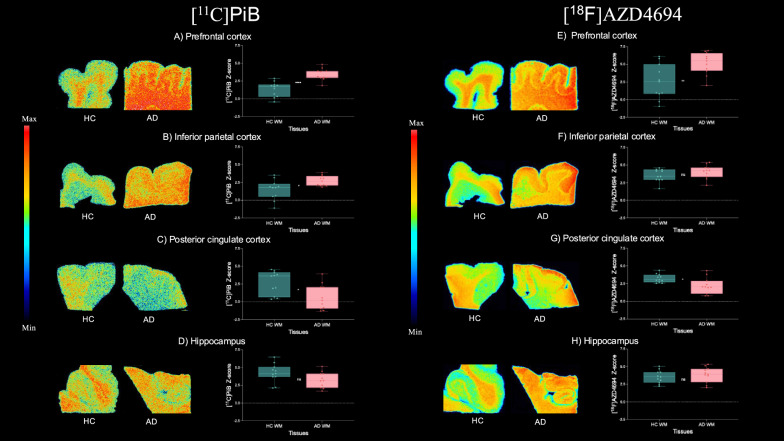
Background: Amyloid-β imaging through positron emission tomography (PET) has significantly transformed Alzheimer's disease (AD) research. [11C]PiB has been widely used for imaging β-amyloid plaques due to its high affinity and selectivity for amyloid deposits. [18F]AZD4694 is a more recently developed amyloid-PET imaging agent, which structurally resembles PiB and has less non-specific binding in the white matter than other 18F-labeled compounds. The purpose of this study is to compare the in vitro binding properties of the amyloid-PET radiotracers [11C]PiB and [18F]AZD4694 in post-mortem human brain tissue. Total binding was assessed by autoradiography in prefrontal, inferior parietal, posterior cingulate cortices and hippocampal sections of healthy control (HC) and AD autopsy-confirmed brain tissues. Furthermore, the displacement of [18F]AZD4694 by unlabeled PiB was evaluated in the above-mentioned sections of AD brain tissues.
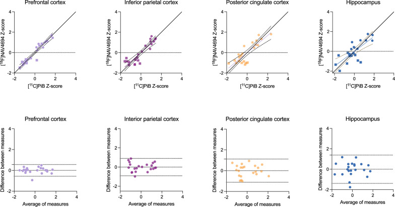
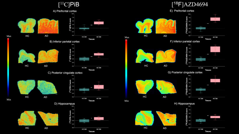
Results: For both radiotracers, we found significant differences (p < 0.0001) between HC and AD tissues binding in the prefrontal cortex ([11C]PiB Cohen's d = 3.424, [18F]AZD4694 Cohen's d = 5.070), inferior parietal cortex ([11C]PiB Cohen's d = 3.156, [18F]AZD4694 Cohen's d = 3.959), posterior cingulate cortex ([11C]PiB Cohen's d = 1.781, [18F]AZD4694 Cohen's d = 3.434), and hippocampus ([11C]PiB Cohen's d = 1.320, [18F]AZD4694 Cohen's d = 3.696). Higher binding was detected for [18F]AZD4694 compared to [11C]PiB in AD prefrontal, inferior parietal and posterior cingulate cortices, while binding in the hippocampus was comparable for both radioligands. Strong correlations between [18]AZD4694 and [11C]PiB were found in the prefrontal (R = 0.959, p < 0.0001), inferior parietal (R = 0.893, p < 0.0001), posterior cingulate (R = 0.838, p = 0.0006) cortices and hippocampus (R = 0.750, p < 0.0001). Bland-Altman analyses revealed strong agreement between [11C]PiB and [18F]AZD4694 in the prefrontal, inferior parietal, and posterior cingulate cortices, but lower agreement in the hippocampus. Displacement studies confirmed high binding affinity of PiB in all tissues, indicating that both amyloid-PET agents compete for the same binding sites.
Conclusions: This head-to-head study provides evidence that while [18F]AZD4694 and [11C]PiB bindings are highly correlated with both tracers competing for the same binding sites, [18F]AZD4694 has a slightly higher effect size when comparing between neuropathologically-confirmed AD and HC brain tissues.
背景:通过正电子发射断层扫描(PET)成像淀粉样蛋白β显著改变了阿尔茨海默病(AD)的研究。[11C]由于PiB对淀粉样蛋白沉积物具有高亲和力和选择性,因此被广泛用于β-淀粉样斑块成像。[18F]AZD4694是最近开发的淀粉样蛋白pet显像剂,其结构类似于PiB,与其他18F标记的化合物相比,在白质中的非特异性结合较少。本研究的目的是比较淀粉样蛋白- pet放射性示踪剂[11C]PiB和[18F]AZD4694在死后人脑组织中的体外结合特性。在健康对照(HC)和AD尸检证实的脑组织的前额叶、下顶叶、后扣带皮层和海马切片中,通过放射自成像评估总结合。此外,在上述AD脑组织切片中,我们评估了未标记PiB对[18F]AZD4694的位移。结果:对于两种放射性示踪剂,我们发现有显著差异(p 11C]PiB Cohen's d = 3.424, [18F]AZD4694 Cohen's d = 5.070),下顶叶皮层([11C]PiB Cohen's d = 3.156, [18F]AZD4694 Cohen's d = 3.959),后扣带皮层([11C]PiB Cohen's d = 1.781, [18F]AZD4694 Cohen's d = 3.434),海马([11C]PiB Cohen's d = 1.320, [18F]AZD4694 Cohen's d = 3.696)。与[11C]PiB相比,[18F]AZD4694在AD前额叶、下顶叶和后扣带皮层的结合程度更高,而两种放射性配体在海马中的结合程度相似。[18]AZD4694与[11C]PiB在前额叶(R = 0.959, p 11C]PiB和[18F]AZD4694在前额叶、下顶叶和后扣带皮层)呈较强相关性,而在海马区呈较低相关性。置换研究证实了PiB在所有组织中的高结合亲和力,表明两种淀粉样蛋白- pet制剂竞争相同的结合位点。结论:这项头对头的研究提供了证据,虽然[18F]AZD4694和[11C]PiB结合与两种示踪剂竞争相同的结合位点高度相关,但[18F]AZD4694在神经病理证实的AD和HC脑组织中具有稍高的效应量。
EJNMMI ResearchRADIOLOGY, NUCLEAR MEDICINE & MEDICAL IMAGING&nb-
CiteScore
5.90
自引率
3.10%
发文量
72
审稿时长
13 weeks
期刊介绍:
EJNMMI Research publishes new basic, translational and clinical research in the field of nuclear medicine and molecular imaging. Regular features include original research articles, rapid communication of preliminary data on innovative research, interesting case reports, editorials, and letters to the editor. Educational articles on basic sciences, fundamental aspects and controversy related to pre-clinical and clinical research or ethical aspects of research are also welcome. Timely reviews provide updates on current applications, issues in imaging research and translational aspects of nuclear medicine and molecular imaging technologies.
The main emphasis is placed on the development of targeted imaging with radiopharmaceuticals within the broader context of molecular probes to enhance understanding and characterisation of the complex biological processes underlying disease and to develop, test and guide new treatment modalities, including radionuclide therapy.

 求助内容:
求助内容: 应助结果提醒方式:


