3'-deoxy-3'-18F-fluorothymidine PET imaging of lymphoid tissues in patients with advanced non-small cell lung cancer undergoing anti-programmed cell death-1 therapy.
IF 3.1 3区 医学Q1 RADIOLOGY, NUCLEAR MEDICINE & MEDICAL IMAGING
{"title":"3'-deoxy-3'-<sup>18</sup>F-fluorothymidine PET imaging of lymphoid tissues in patients with advanced non-small cell lung cancer undergoing anti-programmed cell death-1 therapy.","authors":"Masayuki Sato, Yukihiro Umeda, Tetsuya Tsujikawa, Tetsuya Mori, Akikazu Shimada, Tomoaki Sonoda, Makiko Yamaguchi, Chisato Honjo, Yuko Waseda, Yasushi Kiyono, Tamotsu Ishizuka, Hidehiko Okazawa","doi":"10.1186/s13550-025-01225-7","DOIUrl":null,"url":null,"abstract":"<p><strong>Background: </strong>Anti-programmed cell death-1 (anti-PD-1) therapy has become the standard immunotherapy for patients with advanced non-small cell lung cancer (NSCLC). However, little is known about the organs influenced by PD-1 inhibitors on a patient's tumor immunity. We examined the changes in lymphoid tissue proliferation before and after PD-1 inhibitor treatment using 3'-deoxy-3'-[<sup>18</sup>F]-fluorothymidine (<sup>18</sup>F-FLT) positron emission tomography (PET). This study included 25 patients with advanced NSCLC who underwent <sup>18</sup>F-FLT PET before and 2 and 6 weeks after the initiation of PD-1 inhibitor treatment. We determined the average standardized uptake value (SUV<sub>mean</sub>) in the spleen, maximum SUV (SUV<sub>max</sub>) in the lymph nodes, and the SUV<sub>max</sub>, SUV<sub>mean</sub>, proliferative vertebral volume (PVV), and total vertebral proliferation (TVP) in the thoracolumbar vertebral bodies using <sup>18</sup>F-FLT PET and blood test data. The relationship between the rate of change in these parameters before and after treatment and the tumor response was evaluated.</p><p><strong>Results: </strong>The baseline <sup>18</sup>F-FLT accumulation in the lymphoid tissues or blood test data between the progressive disease (PD) and non-PD groups were not significantly different. In the spleen and lymph nodes, changes in <sup>18</sup>F-FLT accumulation from baseline to 2 or 6 weeks did not differ between the non-PD and PD groups. However, mediastinal lymph node accumulation tended to increase transiently at week 2 compared to that before treatment initiation (median SUV<sub>max</sub> 2.19 vs. 2.64, P = 0.073). Regarding changes in vertebral accumulation in the non-PD group, the SUV<sub>max</sub>, and PVV were significantly lower at weeks 2 and 6. In the percent changes in <sup>18</sup>F-FLT accumulation of the vertebrae after the treatment initiation, the PD group was significantly higher than the non-PD group at the 6-week evaluation (median ΔTVP0-6, 17.0% vs. -13.0%, P = 0.0080).</p><p><strong>Conclusions: </strong>In patients with advanced NSCLC who achieved a tumor response, proliferation decreased in the bone marrow, but not in the spleen or lymph nodes, 6 weeks after treatment initiation. <sup>18</sup>F-FLT PET can help monitor changes in tumor immunity in each lymphoid tissue and may serve as a biomarker for the response to immune checkpoint inhibitor therapy.</p>","PeriodicalId":11611,"journal":{"name":"EJNMMI Research","volume":"15 1","pages":"32"},"PeriodicalIF":3.1000,"publicationDate":"2025-04-01","publicationTypes":"Journal Article","fieldsOfStudy":null,"isOpenAccess":false,"openAccessPdf":"https://www.ncbi.nlm.nih.gov/pmc/articles/PMC11961847/pdf/","citationCount":"0","resultStr":null,"platform":"Semanticscholar","paperid":null,"PeriodicalName":"EJNMMI Research","FirstCategoryId":"3","ListUrlMain":"https://doi.org/10.1186/s13550-025-01225-7","RegionNum":3,"RegionCategory":"医学","ArticlePicture":[],"TitleCN":null,"AbstractTextCN":null,"PMCID":null,"EPubDate":"","PubModel":"","JCR":"Q1","JCRName":"RADIOLOGY, NUCLEAR MEDICINE & MEDICAL IMAGING","Score":null,"Total":0}
引用次数: 0
Abstract
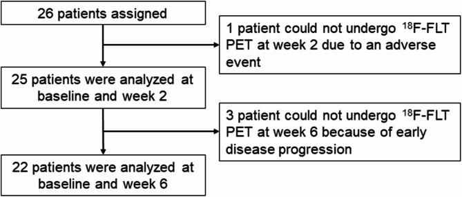
Background: Anti-programmed cell death-1 (anti-PD-1) therapy has become the standard immunotherapy for patients with advanced non-small cell lung cancer (NSCLC). However, little is known about the organs influenced by PD-1 inhibitors on a patient's tumor immunity. We examined the changes in lymphoid tissue proliferation before and after PD-1 inhibitor treatment using 3'-deoxy-3'-[18F]-fluorothymidine (18F-FLT) positron emission tomography (PET). This study included 25 patients with advanced NSCLC who underwent 18F-FLT PET before and 2 and 6 weeks after the initiation of PD-1 inhibitor treatment. We determined the average standardized uptake value (SUVmean) in the spleen, maximum SUV (SUVmax) in the lymph nodes, and the SUVmax, SUVmean, proliferative vertebral volume (PVV), and total vertebral proliferation (TVP) in the thoracolumbar vertebral bodies using 18F-FLT PET and blood test data. The relationship between the rate of change in these parameters before and after treatment and the tumor response was evaluated.
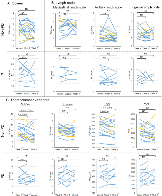
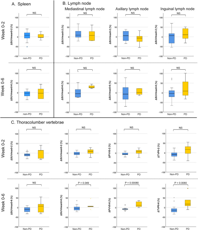
Results: The baseline 18F-FLT accumulation in the lymphoid tissues or blood test data between the progressive disease (PD) and non-PD groups were not significantly different. In the spleen and lymph nodes, changes in 18F-FLT accumulation from baseline to 2 or 6 weeks did not differ between the non-PD and PD groups. However, mediastinal lymph node accumulation tended to increase transiently at week 2 compared to that before treatment initiation (median SUVmax 2.19 vs. 2.64, P = 0.073). Regarding changes in vertebral accumulation in the non-PD group, the SUVmax, and PVV were significantly lower at weeks 2 and 6. In the percent changes in 18F-FLT accumulation of the vertebrae after the treatment initiation, the PD group was significantly higher than the non-PD group at the 6-week evaluation (median ΔTVP0-6, 17.0% vs. -13.0%, P = 0.0080).
Conclusions: In patients with advanced NSCLC who achieved a tumor response, proliferation decreased in the bone marrow, but not in the spleen or lymph nodes, 6 weeks after treatment initiation. 18F-FLT PET can help monitor changes in tumor immunity in each lymphoid tissue and may serve as a biomarker for the response to immune checkpoint inhibitor therapy.
背景:抗程序性细胞死亡-1 (anti-PD-1)治疗已成为晚期非小细胞肺癌(NSCLC)患者的标准免疫治疗方案。然而,PD-1抑制剂对患者肿瘤免疫影响的器官知之甚少。我们使用3'-脱氧-3'-[18F]-氟胸苷(18F- flt)正电子发射断层扫描(PET)检查PD-1抑制剂治疗前后淋巴组织增殖的变化。该研究纳入了25例晚期NSCLC患者,他们在PD-1抑制剂治疗开始前和开始治疗后2周和6周接受了18F-FLT PET。我们使用18F-FLT PET和血液测试数据确定了脾脏的平均标准化摄取值(SUVmean),淋巴结的最大SUV (SUVmax),以及胸腰椎椎体的SUVmax, SUVmean,增生性椎体体积(PVV)和总椎体增殖(TVP)。评估治疗前后这些参数的变化率与肿瘤反应之间的关系。结果:在进展性疾病(PD)组和非PD组之间,淋巴组织中18F-FLT的基线积累或血液检查数据无显著差异。在脾脏和淋巴结中,从基线到2或6周18F-FLT积累的变化在非PD组和PD组之间没有差异。然而,与治疗开始前相比,纵隔淋巴结积聚在第2周有短暂增加的趋势(中位SUVmax 2.19 vs. 2.64, P = 0.073)。关于非pd组椎体堆积的变化,SUVmax和PVV在第2周和第6周显著降低。在治疗开始后椎体18F-FLT积累的百分比变化中,PD组在6周评估时显著高于非PD组(中位数ΔTVP0-6, 17.0% vs. -13.0%, P = 0.0080)。结论:在获得肿瘤应答的晚期NSCLC患者中,在治疗开始6周后,骨髓的增殖减少,但脾脏或淋巴结的增殖没有减少。18F-FLT PET可以帮助监测每个淋巴组织中肿瘤免疫的变化,并可能作为免疫检查点抑制剂治疗反应的生物标志物。
EJNMMI ResearchRADIOLOGY, NUCLEAR MEDICINE & MEDICAL IMAGING&nb-
CiteScore
5.90
自引率
3.10%
发文量
72
审稿时长
13 weeks
期刊介绍:
EJNMMI Research publishes new basic, translational and clinical research in the field of nuclear medicine and molecular imaging. Regular features include original research articles, rapid communication of preliminary data on innovative research, interesting case reports, editorials, and letters to the editor. Educational articles on basic sciences, fundamental aspects and controversy related to pre-clinical and clinical research or ethical aspects of research are also welcome. Timely reviews provide updates on current applications, issues in imaging research and translational aspects of nuclear medicine and molecular imaging technologies.
The main emphasis is placed on the development of targeted imaging with radiopharmaceuticals within the broader context of molecular probes to enhance understanding and characterisation of the complex biological processes underlying disease and to develop, test and guide new treatment modalities, including radionuclide therapy.

 求助内容:
求助内容: 应助结果提醒方式:


