Yuvnik Trada, Mark T Lee, Michael G Jameson, Phillip Chlap, Paul Keall, Daniel Moses, Peter Lin, Allan Fowler
{"title":"Mid-treatment changes in intra-tumoural metabolic heterogeneity correlate to outcomes in oropharyngeal squamous cell carcinoma patients.","authors":"Yuvnik Trada, Mark T Lee, Michael G Jameson, Phillip Chlap, Paul Keall, Daniel Moses, Peter Lin, Allan Fowler","doi":"10.1186/s13550-025-01226-6","DOIUrl":null,"url":null,"abstract":"<p><strong>Background: </strong>This study evaluated mid-treatment changes in intra-tumoural metabolic heterogeneity and quantitative FDG-PET/CT imaging parameters and correlated the changes with treatment outcomes in oropharyngeal squamous cell cancer (OPSCC) patients. 114 patients from two independent cohorts underwent baseline and mid-treatment (week 3) FDG-PET. Standardized uptake value maximum (SUV<sub>max</sub>), standardized uptake value mean (SUV<sub>mean</sub>), metabolic tumour volume (MTV), and total lesional glycolysis (TLG) were measured. Intra-tumoural metabolic heterogeneity was quantified as the area under a cumulative SUV-volume histogram curve (AUC-CSH). Baseline and relative change (%∆) in imaging features were correlated to locoregional recurrence free survival (LRRFS) using multivariate Cox regression analysis. Patients were stratified into three risk groups utilising ∆AUC-CSH and known prognostic features, then compared using Kaplan-Meier analysis.</p><p><strong>Results: </strong>Median follow up was 39 months. 18% of patients developed locoregional recurrence at 2 years. A decrease in heterogeneity (∆AUC-CSH: 24%) was observed mid-treatment. There was no statistically significant difference in tumour heterogeneity (AUC-CSH) at baseline (p = 0.134) and change at week 3 (p = 0.306) between p16 positive and p16 negative patients. Baseline imaging features did not correlate to LRRFS. However, ∆MTV (aHR 1.04; 95% CI 1.03-1.06; p < 0.001) and ∆AUC-CSH (aHR 0.96; 95% CI 0.94-0.98; p = 0.004) were correlated to LRRFS. Stratification using ∆AUC-CSH and p16 status into three groups showed significant differences in LRR (2 year LRRFS 94%, 79%, 17%; log rank p < 0.001). Stratification using ∆AUC-CSH and ∆MTV into three groups showed significant differences in LRR (2 year LRRFS 93%, 70%, 17%; log rank p < 0.001).</p><p><strong>Conclusion: </strong>Mid-treatment changes in intra-tumoural FDG-PET/CT heterogeneity correlated with treatment outcomes in OPSCC and may help with response prediction. These findings suggest potential utility in designing future risk adaptive clinical trials.</p>","PeriodicalId":11611,"journal":{"name":"EJNMMI Research","volume":"15 1","pages":"31"},"PeriodicalIF":3.1000,"publicationDate":"2025-04-01","publicationTypes":"Journal Article","fieldsOfStudy":null,"isOpenAccess":false,"openAccessPdf":"https://www.ncbi.nlm.nih.gov/pmc/articles/PMC11961835/pdf/","citationCount":"0","resultStr":null,"platform":"Semanticscholar","paperid":null,"PeriodicalName":"EJNMMI Research","FirstCategoryId":"3","ListUrlMain":"https://doi.org/10.1186/s13550-025-01226-6","RegionNum":3,"RegionCategory":"医学","ArticlePicture":[],"TitleCN":null,"AbstractTextCN":null,"PMCID":null,"EPubDate":"","PubModel":"","JCR":"Q1","JCRName":"RADIOLOGY, NUCLEAR MEDICINE & MEDICAL IMAGING","Score":null,"Total":0}
引用次数: 0
Abstract
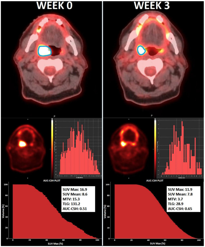
Background: This study evaluated mid-treatment changes in intra-tumoural metabolic heterogeneity and quantitative FDG-PET/CT imaging parameters and correlated the changes with treatment outcomes in oropharyngeal squamous cell cancer (OPSCC) patients. 114 patients from two independent cohorts underwent baseline and mid-treatment (week 3) FDG-PET. Standardized uptake value maximum (SUVmax), standardized uptake value mean (SUVmean), metabolic tumour volume (MTV), and total lesional glycolysis (TLG) were measured. Intra-tumoural metabolic heterogeneity was quantified as the area under a cumulative SUV-volume histogram curve (AUC-CSH). Baseline and relative change (%∆) in imaging features were correlated to locoregional recurrence free survival (LRRFS) using multivariate Cox regression analysis. Patients were stratified into three risk groups utilising ∆AUC-CSH and known prognostic features, then compared using Kaplan-Meier analysis.
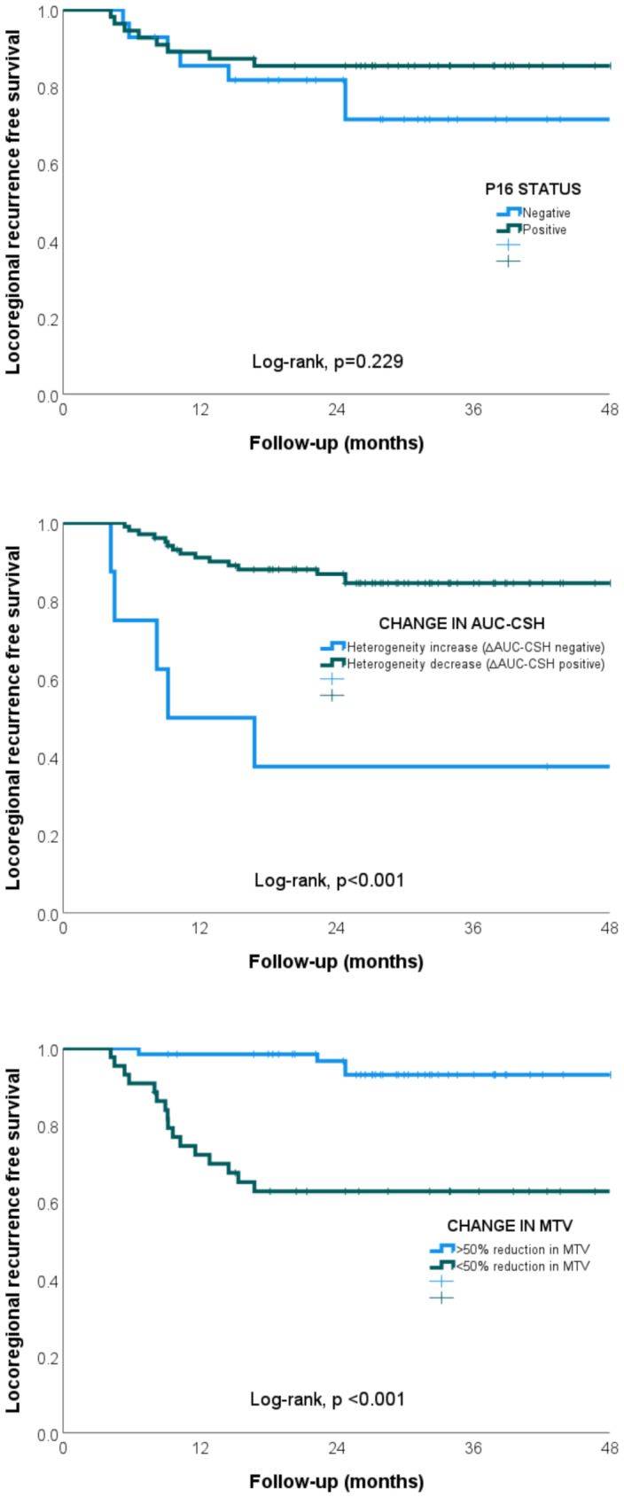
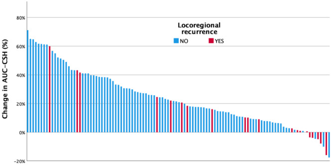
Results: Median follow up was 39 months. 18% of patients developed locoregional recurrence at 2 years. A decrease in heterogeneity (∆AUC-CSH: 24%) was observed mid-treatment. There was no statistically significant difference in tumour heterogeneity (AUC-CSH) at baseline (p = 0.134) and change at week 3 (p = 0.306) between p16 positive and p16 negative patients. Baseline imaging features did not correlate to LRRFS. However, ∆MTV (aHR 1.04; 95% CI 1.03-1.06; p < 0.001) and ∆AUC-CSH (aHR 0.96; 95% CI 0.94-0.98; p = 0.004) were correlated to LRRFS. Stratification using ∆AUC-CSH and p16 status into three groups showed significant differences in LRR (2 year LRRFS 94%, 79%, 17%; log rank p < 0.001). Stratification using ∆AUC-CSH and ∆MTV into three groups showed significant differences in LRR (2 year LRRFS 93%, 70%, 17%; log rank p < 0.001).
Conclusion: Mid-treatment changes in intra-tumoural FDG-PET/CT heterogeneity correlated with treatment outcomes in OPSCC and may help with response prediction. These findings suggest potential utility in designing future risk adaptive clinical trials.
EJNMMI ResearchRADIOLOGY, NUCLEAR MEDICINE & MEDICAL IMAGING&nb-
CiteScore
5.90
自引率
3.10%
发文量
72
审稿时长
13 weeks
期刊介绍:
EJNMMI Research publishes new basic, translational and clinical research in the field of nuclear medicine and molecular imaging. Regular features include original research articles, rapid communication of preliminary data on innovative research, interesting case reports, editorials, and letters to the editor. Educational articles on basic sciences, fundamental aspects and controversy related to pre-clinical and clinical research or ethical aspects of research are also welcome. Timely reviews provide updates on current applications, issues in imaging research and translational aspects of nuclear medicine and molecular imaging technologies.
The main emphasis is placed on the development of targeted imaging with radiopharmaceuticals within the broader context of molecular probes to enhance understanding and characterisation of the complex biological processes underlying disease and to develop, test and guide new treatment modalities, including radionuclide therapy.

 求助内容:
求助内容: 应助结果提醒方式:


