Nicolò Gennaro, Moataz Soliman, Amir A Borhani, Linda Kelahan, Hatice Savas, Ryan Avery, Kamal Subedi, Tugce A Trabzonlu, Chase Krumpelman, Vahid Yaghmai, Young Chae, Jochen Lorch, Devalingam Mahalingam, Mary Mulcahy, Al Benson, Ulas Bagci, Yuri S Velichko
{"title":"Delta Radiomics and Tumor Size: A New Predictive Radiomics Model for Chemotherapy Response in Liver Metastases from Breast and Colorectal Cancer.","authors":"Nicolò Gennaro, Moataz Soliman, Amir A Borhani, Linda Kelahan, Hatice Savas, Ryan Avery, Kamal Subedi, Tugce A Trabzonlu, Chase Krumpelman, Vahid Yaghmai, Young Chae, Jochen Lorch, Devalingam Mahalingam, Mary Mulcahy, Al Benson, Ulas Bagci, Yuri S Velichko","doi":"10.3390/tomography11030020","DOIUrl":null,"url":null,"abstract":"<p><p><b>Background/Objectives</b>: Radiomic features exhibit a correlation with tumor size on pretreatment images. However, on post-treatment images, this association is influenced by treatment efficacy and varies between responders and non-responders. This study introduces a novel model, called baseline-referenced Delta radiomics, which integrates the association between radiomic features and tumor size into Delta radiomics to predict chemotherapy response in liver metastases from breast cancer (BC) and colorectal cancer (CRC). <b>Materials and Methods</b>: A retrospective study analyzed contrast-enhanced computed tomography (CT) scans of 83 BC patients and 84 CRC patients. Among these, 57 BC patients with 106 liver lesions and 37 CRC patients with 109 lesions underwent post-treatment imaging after systemic chemotherapy. Radiomic features were extracted from up to three lesions per patient following manual segmentation. Tumor response was assessed by measuring the longest diameter and classified according to RECIST 1.1 criteria as progressive disease (PD), partial response (PR), or stable disease (SD). Classification models were developed to predict chemotherapy response using pretreatment data only, Delta radiomics, and baseline-referenced Delta radiomics. Model performance was evaluated using confusion matrix metrics. <b>Results</b>: Baseline-referenced Delta radiomics performed comparably or better than established radiomics models in predicting tumor response in chemotherapy-treated patients with liver metastases. The sensitivity, specificity, and balanced accuracy in predicting response ranged from 0.66 to 0.97, 0.81 to 0.97, and 80% to 90%, respectively. <b>Conclusions</b>: By integrating the relationship between radiomic features and tumor size into Delta radiomics, baseline-referenced Delta radiomics offers a promising approach for predicting chemotherapy response in liver metastases from breast and colorectal cancer.</p>","PeriodicalId":51330,"journal":{"name":"Tomography","volume":"11 3","pages":""},"PeriodicalIF":2.2000,"publicationDate":"2025-02-20","publicationTypes":"Journal Article","fieldsOfStudy":null,"isOpenAccess":false,"openAccessPdf":"https://www.ncbi.nlm.nih.gov/pmc/articles/PMC11945686/pdf/","citationCount":"0","resultStr":null,"platform":"Semanticscholar","paperid":null,"PeriodicalName":"Tomography","FirstCategoryId":"3","ListUrlMain":"https://doi.org/10.3390/tomography11030020","RegionNum":4,"RegionCategory":"医学","ArticlePicture":[],"TitleCN":null,"AbstractTextCN":null,"PMCID":null,"EPubDate":"","PubModel":"","JCR":"Q2","JCRName":"RADIOLOGY, NUCLEAR MEDICINE & MEDICAL IMAGING","Score":null,"Total":0}
引用次数: 0
Abstract
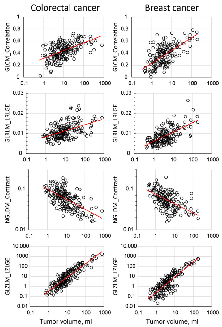
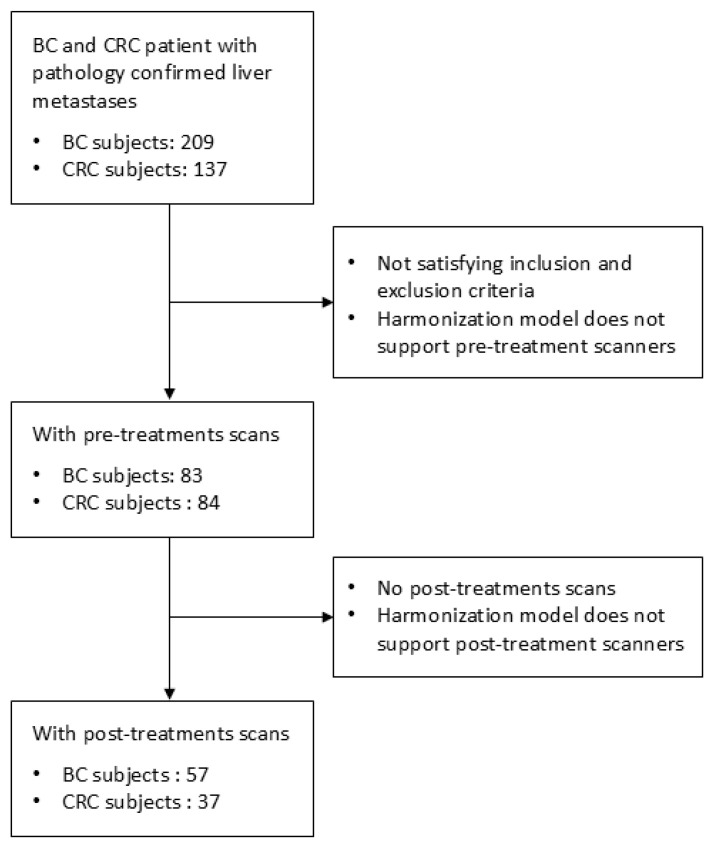
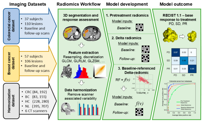
Background/Objectives: Radiomic features exhibit a correlation with tumor size on pretreatment images. However, on post-treatment images, this association is influenced by treatment efficacy and varies between responders and non-responders. This study introduces a novel model, called baseline-referenced Delta radiomics, which integrates the association between radiomic features and tumor size into Delta radiomics to predict chemotherapy response in liver metastases from breast cancer (BC) and colorectal cancer (CRC). Materials and Methods: A retrospective study analyzed contrast-enhanced computed tomography (CT) scans of 83 BC patients and 84 CRC patients. Among these, 57 BC patients with 106 liver lesions and 37 CRC patients with 109 lesions underwent post-treatment imaging after systemic chemotherapy. Radiomic features were extracted from up to three lesions per patient following manual segmentation. Tumor response was assessed by measuring the longest diameter and classified according to RECIST 1.1 criteria as progressive disease (PD), partial response (PR), or stable disease (SD). Classification models were developed to predict chemotherapy response using pretreatment data only, Delta radiomics, and baseline-referenced Delta radiomics. Model performance was evaluated using confusion matrix metrics. Results: Baseline-referenced Delta radiomics performed comparably or better than established radiomics models in predicting tumor response in chemotherapy-treated patients with liver metastases. The sensitivity, specificity, and balanced accuracy in predicting response ranged from 0.66 to 0.97, 0.81 to 0.97, and 80% to 90%, respectively. Conclusions: By integrating the relationship between radiomic features and tumor size into Delta radiomics, baseline-referenced Delta radiomics offers a promising approach for predicting chemotherapy response in liver metastases from breast and colorectal cancer.
TomographyMedicine-Radiology, Nuclear Medicine and Imaging
CiteScore
2.70
自引率
10.50%
发文量
222
期刊介绍:
TomographyTM publishes basic (technical and pre-clinical) and clinical scientific articles which involve the advancement of imaging technologies. Tomography encompasses studies that use single or multiple imaging modalities including for example CT, US, PET, SPECT, MR and hyperpolarization technologies, as well as optical modalities (i.e. bioluminescence, photoacoustic, endomicroscopy, fiber optic imaging and optical computed tomography) in basic sciences, engineering, preclinical and clinical medicine.
Tomography also welcomes studies involving exploration and refinement of contrast mechanisms and image-derived metrics within and across modalities toward the development of novel imaging probes for image-based feedback and intervention. The use of imaging in biology and medicine provides unparalleled opportunities to noninvasively interrogate tissues to obtain real-time dynamic and quantitative information required for diagnosis and response to interventions and to follow evolving pathological conditions. As multi-modal studies and the complexities of imaging technologies themselves are ever increasing to provide advanced information to scientists and clinicians.
Tomography provides a unique publication venue allowing investigators the opportunity to more precisely communicate integrated findings related to the diverse and heterogeneous features associated with underlying anatomical, physiological, functional, metabolic and molecular genetic activities of normal and diseased tissue. Thus Tomography publishes peer-reviewed articles which involve the broad use of imaging of any tissue and disease type including both preclinical and clinical investigations. In addition, hardware/software along with chemical and molecular probe advances are welcome as they are deemed to significantly contribute towards the long-term goal of improving the overall impact of imaging on scientific and clinical discovery.

 求助内容:
求助内容: 应助结果提醒方式:


