Julia Lasek, Karolina Nurzynska, Adam Piórkowski, Michał Strzelecki, Rafał Obuchowicz
{"title":"Deep Learning for Ultrasonographic Assessment of Temporomandibular Joint Morphology.","authors":"Julia Lasek, Karolina Nurzynska, Adam Piórkowski, Michał Strzelecki, Rafał Obuchowicz","doi":"10.3390/tomography11030027","DOIUrl":null,"url":null,"abstract":"<p><strong>Background: </strong>Temporomandibular joint (TMJ) disorders are a significant cause of orofacial pain. Artificial intelligence (AI) has been successfully applied to other imaging modalities but remains underexplored in ultrasonographic evaluations of TMJ.</p><p><strong>Objective: </strong>This study aimed to develop and validate an AI-driven method for the automatic and reproducible measurement of TMJ space width from ultrasonographic images.</p><p><strong>Methods: </strong>A total of 142 TMJ ultrasonographic images were segmented into three anatomical components: the mandibular condyle, joint space, and glenoid fossa. State-of-the-art architectures were tested, and the best-performing 2D Residual U-Net was trained and validated against expert annotations. The algorithm for joint space width measurement based on TMJ segmentation was proposed, calculating the vertical distance between the superior-most point of the mandibular condyle and its corresponding point on the glenoid fossa.</p><p><strong>Results: </strong>The segmentation model achieved high performance for the mandibular condyle (Dice: 0.91 ± 0.08) and joint space (Dice: 0.86 ± 0.09), with notably lower performance for the glenoid fossa (Dice: 0.60 ± 0.24), highlighting variability due to its complex geometry. The TMJ space width measurement algorithm demonstrated minimal bias, with a mean difference of 0.08 mm and a mean absolute error of 0.18 mm compared to reference measurements.</p><p><strong>Conclusions: </strong>The model exhibited potential as a reliable tool for clinical use, demonstrating accuracy in TMJ ultrasonographic analysis. This study underscores the ability of AI-driven segmentation and measurement algorithms to bridge existing gaps in ultrasonographic imaging and lays the foundation for broader clinical applications.</p>","PeriodicalId":51330,"journal":{"name":"Tomography","volume":"11 3","pages":""},"PeriodicalIF":2.2000,"publicationDate":"2025-02-27","publicationTypes":"Journal Article","fieldsOfStudy":null,"isOpenAccess":false,"openAccessPdf":"https://www.ncbi.nlm.nih.gov/pmc/articles/PMC11946603/pdf/","citationCount":"0","resultStr":null,"platform":"Semanticscholar","paperid":null,"PeriodicalName":"Tomography","FirstCategoryId":"3","ListUrlMain":"https://doi.org/10.3390/tomography11030027","RegionNum":4,"RegionCategory":"医学","ArticlePicture":[],"TitleCN":null,"AbstractTextCN":null,"PMCID":null,"EPubDate":"","PubModel":"","JCR":"Q2","JCRName":"RADIOLOGY, NUCLEAR MEDICINE & MEDICAL IMAGING","Score":null,"Total":0}
引用次数: 0
Abstract
Background: Temporomandibular joint (TMJ) disorders are a significant cause of orofacial pain. Artificial intelligence (AI) has been successfully applied to other imaging modalities but remains underexplored in ultrasonographic evaluations of TMJ.
Objective: This study aimed to develop and validate an AI-driven method for the automatic and reproducible measurement of TMJ space width from ultrasonographic images.
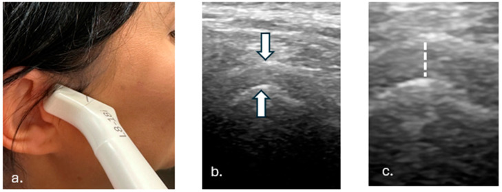
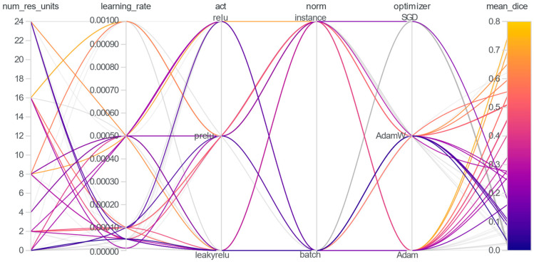
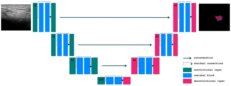
Methods: A total of 142 TMJ ultrasonographic images were segmented into three anatomical components: the mandibular condyle, joint space, and glenoid fossa. State-of-the-art architectures were tested, and the best-performing 2D Residual U-Net was trained and validated against expert annotations. The algorithm for joint space width measurement based on TMJ segmentation was proposed, calculating the vertical distance between the superior-most point of the mandibular condyle and its corresponding point on the glenoid fossa.
Results: The segmentation model achieved high performance for the mandibular condyle (Dice: 0.91 ± 0.08) and joint space (Dice: 0.86 ± 0.09), with notably lower performance for the glenoid fossa (Dice: 0.60 ± 0.24), highlighting variability due to its complex geometry. The TMJ space width measurement algorithm demonstrated minimal bias, with a mean difference of 0.08 mm and a mean absolute error of 0.18 mm compared to reference measurements.
Conclusions: The model exhibited potential as a reliable tool for clinical use, demonstrating accuracy in TMJ ultrasonographic analysis. This study underscores the ability of AI-driven segmentation and measurement algorithms to bridge existing gaps in ultrasonographic imaging and lays the foundation for broader clinical applications.
TomographyMedicine-Radiology, Nuclear Medicine and Imaging
CiteScore
2.70
自引率
10.50%
发文量
222
期刊介绍:
TomographyTM publishes basic (technical and pre-clinical) and clinical scientific articles which involve the advancement of imaging technologies. Tomography encompasses studies that use single or multiple imaging modalities including for example CT, US, PET, SPECT, MR and hyperpolarization technologies, as well as optical modalities (i.e. bioluminescence, photoacoustic, endomicroscopy, fiber optic imaging and optical computed tomography) in basic sciences, engineering, preclinical and clinical medicine.
Tomography also welcomes studies involving exploration and refinement of contrast mechanisms and image-derived metrics within and across modalities toward the development of novel imaging probes for image-based feedback and intervention. The use of imaging in biology and medicine provides unparalleled opportunities to noninvasively interrogate tissues to obtain real-time dynamic and quantitative information required for diagnosis and response to interventions and to follow evolving pathological conditions. As multi-modal studies and the complexities of imaging technologies themselves are ever increasing to provide advanced information to scientists and clinicians.
Tomography provides a unique publication venue allowing investigators the opportunity to more precisely communicate integrated findings related to the diverse and heterogeneous features associated with underlying anatomical, physiological, functional, metabolic and molecular genetic activities of normal and diseased tissue. Thus Tomography publishes peer-reviewed articles which involve the broad use of imaging of any tissue and disease type including both preclinical and clinical investigations. In addition, hardware/software along with chemical and molecular probe advances are welcome as they are deemed to significantly contribute towards the long-term goal of improving the overall impact of imaging on scientific and clinical discovery.

 求助内容:
求助内容: 应助结果提醒方式:


