Siddharth Guha, Abdalla Ibrahim, Pengfei Geng, Qian Wu, Yen Chou, Oguz Akin, Lawrence H Schwartz, Chuan-Miao Xie, Binsheng Zhao
{"title":"Variability of HCC Tumor Diameter and Density Measurements on Dynamic Contrast-Enhanced Computed Tomography.","authors":"Siddharth Guha, Abdalla Ibrahim, Pengfei Geng, Qian Wu, Yen Chou, Oguz Akin, Lawrence H Schwartz, Chuan-Miao Xie, Binsheng Zhao","doi":"10.3390/tomography11030036","DOIUrl":null,"url":null,"abstract":"<p><strong>Purpose: </strong>In cancers imaged using contrast-enhanced protocols, such as hepatocellular carcinoma (HCC), formal guidelines rely on measurements of lesion size (in mm) and radiographic density (in Hounsfield units [HU]) to evaluate response to treatment. However, the variability of these measurements across different contrast enhancement phases remains poorly understood. This limits the ability of clinicians to discern whether measurement changes are accurate.</p><p><strong>Methods: </strong>In this study, we investigated the variability of maximal lesion diameter and mean lesion density of HCC lesions on CT scans across four different contrast enhancement phases: non-contrast-enhanced phase (NCE), early arterial phase (E-AP), late arterial phase (L-AP), and portal venous phase (PVP). HCC lesions were independently segmented by two expert radiologists. For each pair of a lesion's scan timepoints, one was selected randomly as the baseline measurement and the other as the repeat measurement. Both absolute and relative differences in measurements were calculated, as were the coefficients of variance (CVs). Analysis was further stratified by both contrast enhancement phase and lesion diameter.</p><p><strong>Results: </strong>Lesion diameter was found to have a CV of 5.11% (95% CI: 4.20-6.01%). About a fifth of the measurement's relative changes were greater than 10%. Although there was no significant difference in diameter measurements across different phases, there was a significant negative correlation (R = -0.303, <i>p</i>-value = 0.030) between lesion diameter and percent difference in diameter measurement. Lesion density measurements varied significantly across all phases, with the greatest relative difference of 47% in the late arterial phase and a CV of 22.84% (21.48-24.20%). The overall CV for lesion density measurements was 26.19% (24.66-27.72%).</p><p><strong>Conclusions: </strong>Changes in tumor diameter measurements within 10% may simply be due to variability, and lesion density is highly sensitive to contrast timing. This highlights the importance of paying attention to these two variables when evaluating tumor response in both clinical trials and practice.</p>","PeriodicalId":51330,"journal":{"name":"Tomography","volume":"11 3","pages":""},"PeriodicalIF":2.2000,"publicationDate":"2025-03-19","publicationTypes":"Journal Article","fieldsOfStudy":null,"isOpenAccess":false,"openAccessPdf":"https://www.ncbi.nlm.nih.gov/pmc/articles/PMC11946049/pdf/","citationCount":"0","resultStr":null,"platform":"Semanticscholar","paperid":null,"PeriodicalName":"Tomography","FirstCategoryId":"3","ListUrlMain":"https://doi.org/10.3390/tomography11030036","RegionNum":4,"RegionCategory":"医学","ArticlePicture":[],"TitleCN":null,"AbstractTextCN":null,"PMCID":null,"EPubDate":"","PubModel":"","JCR":"Q2","JCRName":"RADIOLOGY, NUCLEAR MEDICINE & MEDICAL IMAGING","Score":null,"Total":0}
引用次数: 0
Abstract
Purpose: In cancers imaged using contrast-enhanced protocols, such as hepatocellular carcinoma (HCC), formal guidelines rely on measurements of lesion size (in mm) and radiographic density (in Hounsfield units [HU]) to evaluate response to treatment. However, the variability of these measurements across different contrast enhancement phases remains poorly understood. This limits the ability of clinicians to discern whether measurement changes are accurate.
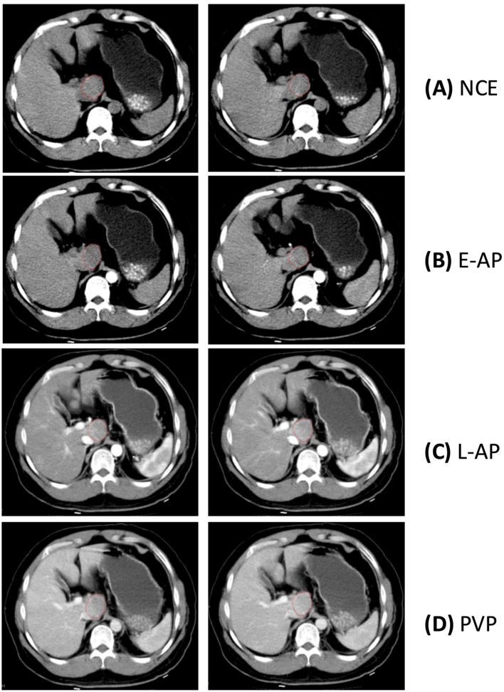
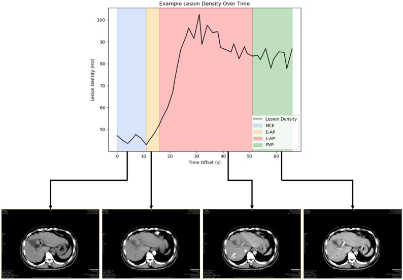
Methods: In this study, we investigated the variability of maximal lesion diameter and mean lesion density of HCC lesions on CT scans across four different contrast enhancement phases: non-contrast-enhanced phase (NCE), early arterial phase (E-AP), late arterial phase (L-AP), and portal venous phase (PVP). HCC lesions were independently segmented by two expert radiologists. For each pair of a lesion's scan timepoints, one was selected randomly as the baseline measurement and the other as the repeat measurement. Both absolute and relative differences in measurements were calculated, as were the coefficients of variance (CVs). Analysis was further stratified by both contrast enhancement phase and lesion diameter.
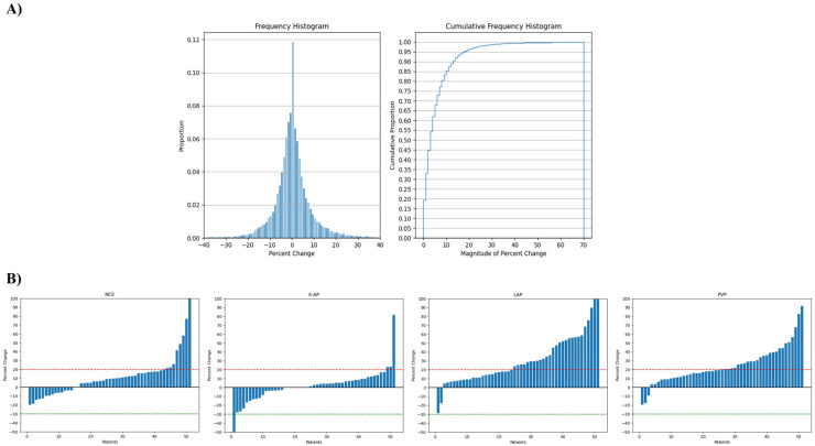
Results: Lesion diameter was found to have a CV of 5.11% (95% CI: 4.20-6.01%). About a fifth of the measurement's relative changes were greater than 10%. Although there was no significant difference in diameter measurements across different phases, there was a significant negative correlation (R = -0.303, p-value = 0.030) between lesion diameter and percent difference in diameter measurement. Lesion density measurements varied significantly across all phases, with the greatest relative difference of 47% in the late arterial phase and a CV of 22.84% (21.48-24.20%). The overall CV for lesion density measurements was 26.19% (24.66-27.72%).
Conclusions: Changes in tumor diameter measurements within 10% may simply be due to variability, and lesion density is highly sensitive to contrast timing. This highlights the importance of paying attention to these two variables when evaluating tumor response in both clinical trials and practice.
TomographyMedicine-Radiology, Nuclear Medicine and Imaging
CiteScore
2.70
自引率
10.50%
发文量
222
期刊介绍:
TomographyTM publishes basic (technical and pre-clinical) and clinical scientific articles which involve the advancement of imaging technologies. Tomography encompasses studies that use single or multiple imaging modalities including for example CT, US, PET, SPECT, MR and hyperpolarization technologies, as well as optical modalities (i.e. bioluminescence, photoacoustic, endomicroscopy, fiber optic imaging and optical computed tomography) in basic sciences, engineering, preclinical and clinical medicine.
Tomography also welcomes studies involving exploration and refinement of contrast mechanisms and image-derived metrics within and across modalities toward the development of novel imaging probes for image-based feedback and intervention. The use of imaging in biology and medicine provides unparalleled opportunities to noninvasively interrogate tissues to obtain real-time dynamic and quantitative information required for diagnosis and response to interventions and to follow evolving pathological conditions. As multi-modal studies and the complexities of imaging technologies themselves are ever increasing to provide advanced information to scientists and clinicians.
Tomography provides a unique publication venue allowing investigators the opportunity to more precisely communicate integrated findings related to the diverse and heterogeneous features associated with underlying anatomical, physiological, functional, metabolic and molecular genetic activities of normal and diseased tissue. Thus Tomography publishes peer-reviewed articles which involve the broad use of imaging of any tissue and disease type including both preclinical and clinical investigations. In addition, hardware/software along with chemical and molecular probe advances are welcome as they are deemed to significantly contribute towards the long-term goal of improving the overall impact of imaging on scientific and clinical discovery.

 求助内容:
求助内容: 应助结果提醒方式:


