Prediction of Chemotherapy Response in Locally Advanced Breast Cancer Patients at Pre-Treatment Using CT Textural Features and Machine Learning: Comparison of Feature Selection Methods.
IF 2.2 4区 医学Q2 RADIOLOGY, NUCLEAR MEDICINE & MEDICAL IMAGING
Amir Moslemi, Laurentius Oscar Osapoetra, Archya Dasgupta, Schontal Halstead, David Alberico, Maureen Trudeau, Sonal Gandhi, Andrea Eisen, Frances Wright, Nicole Look-Hong, Belinda Curpen, Michael Kolios, Gregory J Czarnota
{"title":"Prediction of Chemotherapy Response in Locally Advanced Breast Cancer Patients at Pre-Treatment Using CT Textural Features and Machine Learning: Comparison of Feature Selection Methods.","authors":"Amir Moslemi, Laurentius Oscar Osapoetra, Archya Dasgupta, Schontal Halstead, David Alberico, Maureen Trudeau, Sonal Gandhi, Andrea Eisen, Frances Wright, Nicole Look-Hong, Belinda Curpen, Michael Kolios, Gregory J Czarnota","doi":"10.3390/tomography11030033","DOIUrl":null,"url":null,"abstract":"<p><strong>Rationale: </strong>Neoadjuvant chemotherapy (NAC) is a key element of treatment for locally advanced breast cancer (LABC). Predicting the response of NAC for patients with LABC before initiating treatment would be valuable to customize therapies and ensure the delivery of effective care.</p><p><strong>Objective: </strong>Our objective was to develop predictive measures of tumor response to NAC prior to starting for LABC using machine learning and textural computed tomography (CT) features in different level of frequencies.</p><p><strong>Materials and methods: </strong>A total of 851 textural biomarkers were determined from CT images and their wavelet coefficients for 117 patients with LABC to evaluate the response to NAC. A machine learning pipeline was designed to classify response to NAC treatment for patients with LABC. For training predictive models, three models including all features (wavelet and original image features), only wavelet and only original-image features were considered. We determined features from CT images in different level of frequencies using wavelet transform. Additionally, we conducted a comparison of feature selection methods including mRMR, Relief, Rref QR decomposition, nonnegative matrix factorization and perturbation theory feature selection techniques.</p><p><strong>Results: </strong>Of the 117 patients with LABC evaluated, 82 (70%) had clinical-pathological response to chemotherapy and 35 (30%) had no response to chemotherapy. The best performance for hold-out data splitting was obtained using the KNN classifier using the Top-5 features, which were obtained by mRMR, for all features (accuracy = 77%, specificity = 80%, sensitivity = 56%, and balanced-accuracy = 68%). Likewise, the best performance for leave-one-out data splitting could be obtained by the KNN classifier using the Top-5 features, which was obtained by mRMR, for all features (accuracy = 75%, specificity = 76%, sensitivity = 62%, and balanced-accuracy = 72%).</p><p><strong>Conclusions: </strong>The combination of original textural features and wavelet features results in a greater predictive accuracy of NAC response for LABC patients. This predictive model can be utilized to predict treatment outcomes prior to starting, and clinicians can use it as a recommender system to modify treatment.</p>","PeriodicalId":51330,"journal":{"name":"Tomography","volume":"11 3","pages":""},"PeriodicalIF":2.2000,"publicationDate":"2025-03-13","publicationTypes":"Journal Article","fieldsOfStudy":null,"isOpenAccess":false,"openAccessPdf":"https://www.ncbi.nlm.nih.gov/pmc/articles/PMC11946754/pdf/","citationCount":"0","resultStr":null,"platform":"Semanticscholar","paperid":null,"PeriodicalName":"Tomography","FirstCategoryId":"3","ListUrlMain":"https://doi.org/10.3390/tomography11030033","RegionNum":4,"RegionCategory":"医学","ArticlePicture":[],"TitleCN":null,"AbstractTextCN":null,"PMCID":null,"EPubDate":"","PubModel":"","JCR":"Q2","JCRName":"RADIOLOGY, NUCLEAR MEDICINE & MEDICAL IMAGING","Score":null,"Total":0}
引用次数: 0
Abstract
Rationale: Neoadjuvant chemotherapy (NAC) is a key element of treatment for locally advanced breast cancer (LABC). Predicting the response of NAC for patients with LABC before initiating treatment would be valuable to customize therapies and ensure the delivery of effective care.
Objective: Our objective was to develop predictive measures of tumor response to NAC prior to starting for LABC using machine learning and textural computed tomography (CT) features in different level of frequencies.
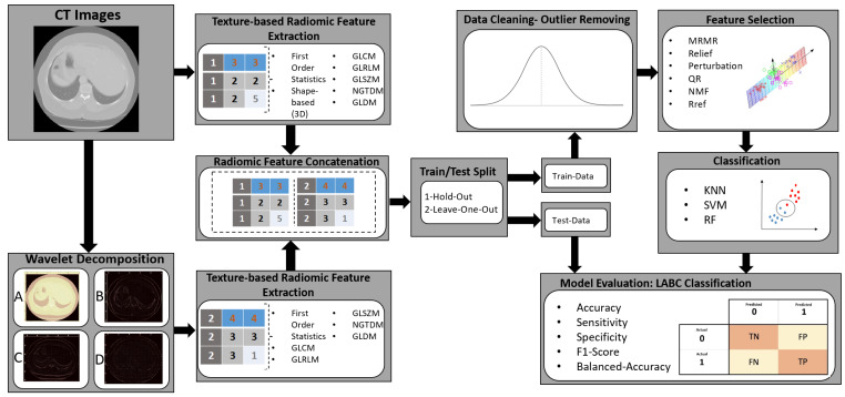
Materials and methods: A total of 851 textural biomarkers were determined from CT images and their wavelet coefficients for 117 patients with LABC to evaluate the response to NAC. A machine learning pipeline was designed to classify response to NAC treatment for patients with LABC. For training predictive models, three models including all features (wavelet and original image features), only wavelet and only original-image features were considered. We determined features from CT images in different level of frequencies using wavelet transform. Additionally, we conducted a comparison of feature selection methods including mRMR, Relief, Rref QR decomposition, nonnegative matrix factorization and perturbation theory feature selection techniques.
Results: Of the 117 patients with LABC evaluated, 82 (70%) had clinical-pathological response to chemotherapy and 35 (30%) had no response to chemotherapy. The best performance for hold-out data splitting was obtained using the KNN classifier using the Top-5 features, which were obtained by mRMR, for all features (accuracy = 77%, specificity = 80%, sensitivity = 56%, and balanced-accuracy = 68%). Likewise, the best performance for leave-one-out data splitting could be obtained by the KNN classifier using the Top-5 features, which was obtained by mRMR, for all features (accuracy = 75%, specificity = 76%, sensitivity = 62%, and balanced-accuracy = 72%).
Conclusions: The combination of original textural features and wavelet features results in a greater predictive accuracy of NAC response for LABC patients. This predictive model can be utilized to predict treatment outcomes prior to starting, and clinicians can use it as a recommender system to modify treatment.
TomographyMedicine-Radiology, Nuclear Medicine and Imaging
CiteScore
2.70
自引率
10.50%
发文量
222
期刊介绍:
TomographyTM publishes basic (technical and pre-clinical) and clinical scientific articles which involve the advancement of imaging technologies. Tomography encompasses studies that use single or multiple imaging modalities including for example CT, US, PET, SPECT, MR and hyperpolarization technologies, as well as optical modalities (i.e. bioluminescence, photoacoustic, endomicroscopy, fiber optic imaging and optical computed tomography) in basic sciences, engineering, preclinical and clinical medicine.
Tomography also welcomes studies involving exploration and refinement of contrast mechanisms and image-derived metrics within and across modalities toward the development of novel imaging probes for image-based feedback and intervention. The use of imaging in biology and medicine provides unparalleled opportunities to noninvasively interrogate tissues to obtain real-time dynamic and quantitative information required for diagnosis and response to interventions and to follow evolving pathological conditions. As multi-modal studies and the complexities of imaging technologies themselves are ever increasing to provide advanced information to scientists and clinicians.
Tomography provides a unique publication venue allowing investigators the opportunity to more precisely communicate integrated findings related to the diverse and heterogeneous features associated with underlying anatomical, physiological, functional, metabolic and molecular genetic activities of normal and diseased tissue. Thus Tomography publishes peer-reviewed articles which involve the broad use of imaging of any tissue and disease type including both preclinical and clinical investigations. In addition, hardware/software along with chemical and molecular probe advances are welcome as they are deemed to significantly contribute towards the long-term goal of improving the overall impact of imaging on scientific and clinical discovery.

 求助内容:
求助内容: 应助结果提醒方式:


