Longitudinal Analysis of Amyloid PET and Brain MRI for Predicting Conversion from Mild Cognitive Impairment to Alzheimer's Disease: Findings from the ADNI Cohort.
IF 2.2 4区 医学Q2 RADIOLOGY, NUCLEAR MEDICINE & MEDICAL IMAGING
{"title":"Longitudinal Analysis of Amyloid PET and Brain MRI for Predicting Conversion from Mild Cognitive Impairment to Alzheimer's Disease: Findings from the ADNI Cohort.","authors":"Do-Hoon Kim","doi":"10.3390/tomography11030037","DOIUrl":null,"url":null,"abstract":"<p><strong>Background/objectives: </strong>This study aimed to investigate the predictive power of integrated longitudinal amyloid positron emission tomography (PET) and brain magnetic resonance imaging (MRI) data for determining the likelihood of conversion to Alzheimer's disease (AD) in patients with mild cognitive impairment (MCI).</p><p><strong>Methods: </strong>We included 180 patients with MCI from the Alzheimer's Disease Neuroimaging Initiative, with baseline and 2-year follow-up scans obtained using F-18 florbetapir PET and MRI. Patients were categorized as converters (progressing to AD) or nonconverters based on a 6-year follow-up. Quantitative analyses included the calculation of amyloid burden using the standardized uptake value ratio (SUVR), brain amyloid smoothing scores (BASSs), brain atrophy indices (BAIs), and their integration into shape features. Longitudinal changes and receiver operating characteristic analyses assessed the predictive power of these biomarkers.</p><p><strong>Results: </strong>Among 180 patients with MCI, 76 (42.2%) were converters, who exhibited significantly higher baseline and 2-year follow-up values for SUVR, BASS, BAI, and shape features than nonconverters (<i>p</i> < 0.001). Shape features demonstrated the highest predictive accuracy for conversion, with areas under the curve of 0.891 at baseline and 0.898 at 2 years. Percent change analyses revealed significant increases in brain atrophy; amyloid deposition changes showed a paradoxical decrease in converters. Additionally, strong associations were observed between longitudinal changes in shape features and neuropsychological test results.</p><p><strong>Conclusions: </strong>The integration of amyloid PET and MRI biomarkers enhances the prediction of AD progression in patients with MCI. These findings support the potential of combined imaging approaches for early diagnosis and targeted interventions in AD.</p>","PeriodicalId":51330,"journal":{"name":"Tomography","volume":"11 3","pages":""},"PeriodicalIF":2.2000,"publicationDate":"2025-03-19","publicationTypes":"Journal Article","fieldsOfStudy":null,"isOpenAccess":false,"openAccessPdf":"https://www.ncbi.nlm.nih.gov/pmc/articles/PMC11945403/pdf/","citationCount":"0","resultStr":null,"platform":"Semanticscholar","paperid":null,"PeriodicalName":"Tomography","FirstCategoryId":"3","ListUrlMain":"https://doi.org/10.3390/tomography11030037","RegionNum":4,"RegionCategory":"医学","ArticlePicture":[],"TitleCN":null,"AbstractTextCN":null,"PMCID":null,"EPubDate":"","PubModel":"","JCR":"Q2","JCRName":"RADIOLOGY, NUCLEAR MEDICINE & MEDICAL IMAGING","Score":null,"Total":0}
引用次数: 0
Abstract
Background/objectives: This study aimed to investigate the predictive power of integrated longitudinal amyloid positron emission tomography (PET) and brain magnetic resonance imaging (MRI) data for determining the likelihood of conversion to Alzheimer's disease (AD) in patients with mild cognitive impairment (MCI).
Methods: We included 180 patients with MCI from the Alzheimer's Disease Neuroimaging Initiative, with baseline and 2-year follow-up scans obtained using F-18 florbetapir PET and MRI. Patients were categorized as converters (progressing to AD) or nonconverters based on a 6-year follow-up. Quantitative analyses included the calculation of amyloid burden using the standardized uptake value ratio (SUVR), brain amyloid smoothing scores (BASSs), brain atrophy indices (BAIs), and their integration into shape features. Longitudinal changes and receiver operating characteristic analyses assessed the predictive power of these biomarkers.
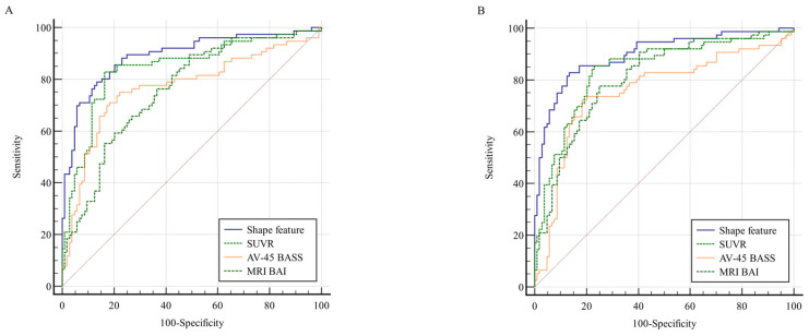
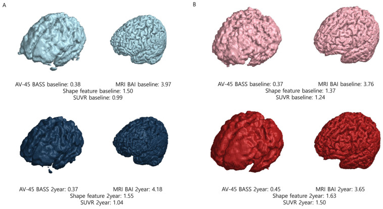
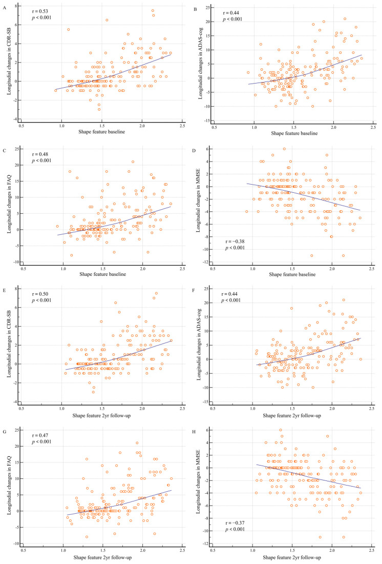
Results: Among 180 patients with MCI, 76 (42.2%) were converters, who exhibited significantly higher baseline and 2-year follow-up values for SUVR, BASS, BAI, and shape features than nonconverters (p < 0.001). Shape features demonstrated the highest predictive accuracy for conversion, with areas under the curve of 0.891 at baseline and 0.898 at 2 years. Percent change analyses revealed significant increases in brain atrophy; amyloid deposition changes showed a paradoxical decrease in converters. Additionally, strong associations were observed between longitudinal changes in shape features and neuropsychological test results.
Conclusions: The integration of amyloid PET and MRI biomarkers enhances the prediction of AD progression in patients with MCI. These findings support the potential of combined imaging approaches for early diagnosis and targeted interventions in AD.
TomographyMedicine-Radiology, Nuclear Medicine and Imaging
CiteScore
2.70
自引率
10.50%
发文量
222
期刊介绍:
TomographyTM publishes basic (technical and pre-clinical) and clinical scientific articles which involve the advancement of imaging technologies. Tomography encompasses studies that use single or multiple imaging modalities including for example CT, US, PET, SPECT, MR and hyperpolarization technologies, as well as optical modalities (i.e. bioluminescence, photoacoustic, endomicroscopy, fiber optic imaging and optical computed tomography) in basic sciences, engineering, preclinical and clinical medicine.
Tomography also welcomes studies involving exploration and refinement of contrast mechanisms and image-derived metrics within and across modalities toward the development of novel imaging probes for image-based feedback and intervention. The use of imaging in biology and medicine provides unparalleled opportunities to noninvasively interrogate tissues to obtain real-time dynamic and quantitative information required for diagnosis and response to interventions and to follow evolving pathological conditions. As multi-modal studies and the complexities of imaging technologies themselves are ever increasing to provide advanced information to scientists and clinicians.
Tomography provides a unique publication venue allowing investigators the opportunity to more precisely communicate integrated findings related to the diverse and heterogeneous features associated with underlying anatomical, physiological, functional, metabolic and molecular genetic activities of normal and diseased tissue. Thus Tomography publishes peer-reviewed articles which involve the broad use of imaging of any tissue and disease type including both preclinical and clinical investigations. In addition, hardware/software along with chemical and molecular probe advances are welcome as they are deemed to significantly contribute towards the long-term goal of improving the overall impact of imaging on scientific and clinical discovery.

 求助内容:
求助内容: 应助结果提醒方式:


