Distinguishing Low Expression Levels of Human Epidermal Growth Factor Receptor 2 in Breast Cancer: Insights from Qualitative and Quantitative Magnetic Resonance Imaging Analysis.
IF 2.2 4区 医学Q2 RADIOLOGY, NUCLEAR MEDICINE & MEDICAL IMAGING
{"title":"Distinguishing Low Expression Levels of Human Epidermal Growth Factor Receptor 2 in Breast Cancer: Insights from Qualitative and Quantitative Magnetic Resonance Imaging Analysis.","authors":"Yiyuan Shen, Xu Zhang, Jinlong Zheng, Simin Wang, Jie Ding, Shiyun Sun, Qianming Bai, Caixia Fu, Junlong Wang, Jing Gong, Chao You, Yajia Gu","doi":"10.3390/tomography11030031","DOIUrl":null,"url":null,"abstract":"<p><strong>Background: </strong>The discovery of novel antibody-drug conjugates for low-expression human epidermal growth factor receptor 2 (HER2-low) breast cancer highlights the inadequacy of the conventional binary classification of HER2 status as either negative or positive. Identification of HER2-low breast cancer is crucial for selecting patients who may benefit from targeted therapies. This study aims to determine whether qualitative and quantitative magnetic resonance imaging (MRI) features can effectively reflect low-HER2-expression breast cancer.</p><p><strong>Methods: </strong>Pre-treatment breast MRI images from 232 patients with pathologically confirmed breast cancer were retrospectively analyzed. Both clinicopathologic and MRI features were recorded. Qualitative MRI features included Breast Imaging Reporting and Data System (BI-RADS) descriptors from dynamic contrast-enhanced MRI (DCE-MRI), as well as intratumoral T2 hyperintensity and peritumoral edema observed in T2-weighted imaging (T2WI). Quantitative features were derived from diffusion kurtosis imaging (DKI) using multiple b-values and included statistics such as mean, median, 5th and 95th percentiles, skewness, kurtosis, and entropy from apparent diffusion coefficient (ADC), D<sub>app</sub>, and K<sub>app</sub> histograms. Differences in clinicopathologic, qualitative, and quantitative MRI features were compared across groups, with multivariable logistic regression used to identify significant independent predictors of HER2-low breast cancer. The discriminative power of MRI features was assessed using receiver operating characteristic (ROC) curves.</p><p><strong>Results: </strong>HER2 status was categorized as HER2-zero (n = 60), HER2-low (n = 91), and HER2-overexpressed (n = 81). Clinically, estrogen receptor (ER), progesterone receptor (PR), hormone receptor (HR), and Ki-67 levels significantly differed between the HER2-low group and others (all <i>p</i> < 0.001). In MRI analyses, intratumoral T2 hyperintensity was more prevalent in HER2-low cases (<i>p</i> = 0.009, <i>p</i> = 0.008). Mass lesions were more common in the HER2-zero group than in the HER2-low group (<i>p</i> = 0.038), and mass shape (<i>p</i> < 0.001) and margin (<i>p</i> < 0.001) significantly varied between the HER2 groups, with mass shape emerging as an independent predictive factor (HER2-low vs. HER2-zero: <i>p</i> = 0.010, HER2-low vs. HER2-over: <i>p</i> = 0.012). Qualitative MRI features demonstrated an area under the curve (AUC) of 0.763 (95% confidence interval [CI]: 0.667-0.859) for distinguishing HER2-low from HER2-zero status. Quantitative features showed distinct differences between HER2-low and HER2-overexpression groups, particularly in non-mass enhancement (NME) lesions. Combined variables achieved the highest predictive accuracy for HER2-low status, with an AUC of 0.802 (95% CI: 0.701-0.903).</p><p><strong>Conclusions: </strong>Qualitative and quantitative MRI features offer valuable insights into low-HER2-expression breast cancer. While qualitative features are more effective for mass lesions, quantitative features are more suitable for NME lesions. These findings provide a more accessible and cost-effective approach to noninvasively identifying patients who may benefit from targeted therapy.</p>","PeriodicalId":51330,"journal":{"name":"Tomography","volume":"11 3","pages":""},"PeriodicalIF":2.2000,"publicationDate":"2025-03-10","publicationTypes":"Journal Article","fieldsOfStudy":null,"isOpenAccess":false,"openAccessPdf":"https://www.ncbi.nlm.nih.gov/pmc/articles/PMC11945706/pdf/","citationCount":"0","resultStr":null,"platform":"Semanticscholar","paperid":null,"PeriodicalName":"Tomography","FirstCategoryId":"3","ListUrlMain":"https://doi.org/10.3390/tomography11030031","RegionNum":4,"RegionCategory":"医学","ArticlePicture":[],"TitleCN":null,"AbstractTextCN":null,"PMCID":null,"EPubDate":"","PubModel":"","JCR":"Q2","JCRName":"RADIOLOGY, NUCLEAR MEDICINE & MEDICAL IMAGING","Score":null,"Total":0}
引用次数: 0
Abstract
Background: The discovery of novel antibody-drug conjugates for low-expression human epidermal growth factor receptor 2 (HER2-low) breast cancer highlights the inadequacy of the conventional binary classification of HER2 status as either negative or positive. Identification of HER2-low breast cancer is crucial for selecting patients who may benefit from targeted therapies. This study aims to determine whether qualitative and quantitative magnetic resonance imaging (MRI) features can effectively reflect low-HER2-expression breast cancer.
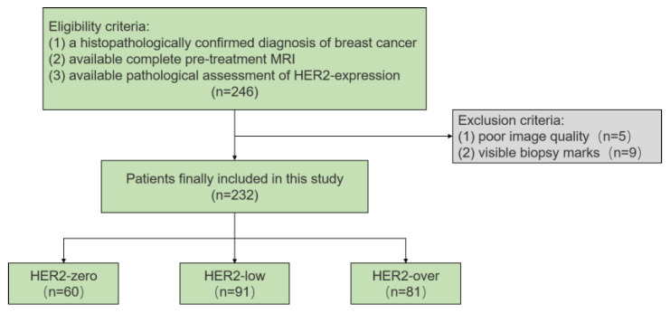
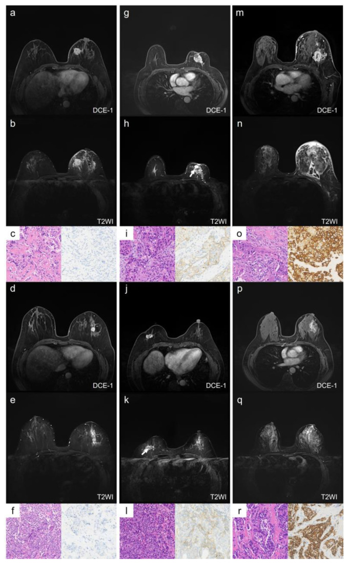
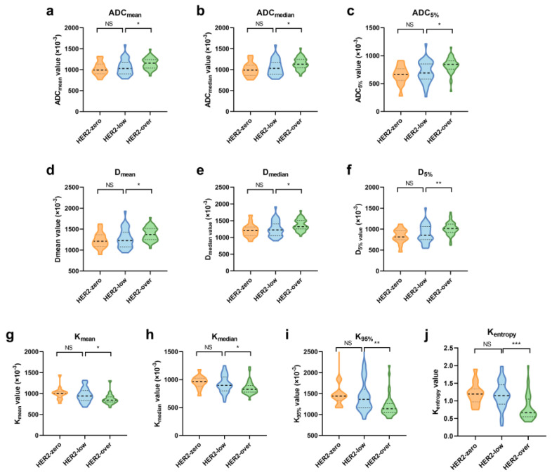
Methods: Pre-treatment breast MRI images from 232 patients with pathologically confirmed breast cancer were retrospectively analyzed. Both clinicopathologic and MRI features were recorded. Qualitative MRI features included Breast Imaging Reporting and Data System (BI-RADS) descriptors from dynamic contrast-enhanced MRI (DCE-MRI), as well as intratumoral T2 hyperintensity and peritumoral edema observed in T2-weighted imaging (T2WI). Quantitative features were derived from diffusion kurtosis imaging (DKI) using multiple b-values and included statistics such as mean, median, 5th and 95th percentiles, skewness, kurtosis, and entropy from apparent diffusion coefficient (ADC), Dapp, and Kapp histograms. Differences in clinicopathologic, qualitative, and quantitative MRI features were compared across groups, with multivariable logistic regression used to identify significant independent predictors of HER2-low breast cancer. The discriminative power of MRI features was assessed using receiver operating characteristic (ROC) curves.
Results: HER2 status was categorized as HER2-zero (n = 60), HER2-low (n = 91), and HER2-overexpressed (n = 81). Clinically, estrogen receptor (ER), progesterone receptor (PR), hormone receptor (HR), and Ki-67 levels significantly differed between the HER2-low group and others (all p < 0.001). In MRI analyses, intratumoral T2 hyperintensity was more prevalent in HER2-low cases (p = 0.009, p = 0.008). Mass lesions were more common in the HER2-zero group than in the HER2-low group (p = 0.038), and mass shape (p < 0.001) and margin (p < 0.001) significantly varied between the HER2 groups, with mass shape emerging as an independent predictive factor (HER2-low vs. HER2-zero: p = 0.010, HER2-low vs. HER2-over: p = 0.012). Qualitative MRI features demonstrated an area under the curve (AUC) of 0.763 (95% confidence interval [CI]: 0.667-0.859) for distinguishing HER2-low from HER2-zero status. Quantitative features showed distinct differences between HER2-low and HER2-overexpression groups, particularly in non-mass enhancement (NME) lesions. Combined variables achieved the highest predictive accuracy for HER2-low status, with an AUC of 0.802 (95% CI: 0.701-0.903).
Conclusions: Qualitative and quantitative MRI features offer valuable insights into low-HER2-expression breast cancer. While qualitative features are more effective for mass lesions, quantitative features are more suitable for NME lesions. These findings provide a more accessible and cost-effective approach to noninvasively identifying patients who may benefit from targeted therapy.
TomographyMedicine-Radiology, Nuclear Medicine and Imaging
CiteScore
2.70
自引率
10.50%
发文量
222
期刊介绍:
TomographyTM publishes basic (technical and pre-clinical) and clinical scientific articles which involve the advancement of imaging technologies. Tomography encompasses studies that use single or multiple imaging modalities including for example CT, US, PET, SPECT, MR and hyperpolarization technologies, as well as optical modalities (i.e. bioluminescence, photoacoustic, endomicroscopy, fiber optic imaging and optical computed tomography) in basic sciences, engineering, preclinical and clinical medicine.
Tomography also welcomes studies involving exploration and refinement of contrast mechanisms and image-derived metrics within and across modalities toward the development of novel imaging probes for image-based feedback and intervention. The use of imaging in biology and medicine provides unparalleled opportunities to noninvasively interrogate tissues to obtain real-time dynamic and quantitative information required for diagnosis and response to interventions and to follow evolving pathological conditions. As multi-modal studies and the complexities of imaging technologies themselves are ever increasing to provide advanced information to scientists and clinicians.
Tomography provides a unique publication venue allowing investigators the opportunity to more precisely communicate integrated findings related to the diverse and heterogeneous features associated with underlying anatomical, physiological, functional, metabolic and molecular genetic activities of normal and diseased tissue. Thus Tomography publishes peer-reviewed articles which involve the broad use of imaging of any tissue and disease type including both preclinical and clinical investigations. In addition, hardware/software along with chemical and molecular probe advances are welcome as they are deemed to significantly contribute towards the long-term goal of improving the overall impact of imaging on scientific and clinical discovery.

 求助内容:
求助内容: 应助结果提醒方式:


