Hannah Hummel-Abmeier, Sabine Naxer, Ella Maria Kadas, Hanna Zimmermann, Bianca Knaack, Peter Huppke, Antonia Kowallick, Kolja Meier, Alexander Ulrich Brandt, Friedemann Paul, Michael Schittkowski, Frederike Cosima Oertel, Jutta Gärtner
{"title":"The Inner Nuclear Layer in Pediatric Multiple Sclerosis.","authors":"Hannah Hummel-Abmeier, Sabine Naxer, Ella Maria Kadas, Hanna Zimmermann, Bianca Knaack, Peter Huppke, Antonia Kowallick, Kolja Meier, Alexander Ulrich Brandt, Friedemann Paul, Michael Schittkowski, Frederike Cosima Oertel, Jutta Gärtner","doi":"10.1212/NXI.0000000000200387","DOIUrl":null,"url":null,"abstract":"<p><strong>Background and objectives: </strong>Pediatric onset multiple sclerosis (POMS) leads to optic nerve and retinal damage from optic neuritis (ON) and potential subclinical disease activity. Neuroaxonal retinal damage manifests in peripapillary retinal nerve fiber layer (pRNFL) and macular ganglion cell and inner plexiform layer (GCIP) thinning. Inner nuclear layer (INL) thickness has been suggested to increase with inflammatory activity or after acute ON, and decrease from chronic neurodegeneration. Macular microcysts in the INL have been described in patients with adult MS. The objective of this study was to investigate the INL in a large cohort of POMS as a potential biomarker for evaluation of disease course and therapeutic success.</p><p><strong>Methods: </strong>For this cross-sectional case-control study, we prospectively recruited 153 patients with POMS and 92 controls, including asymptomatic healthy volunteers and children admitted to the hospital with nonretinal disorders. Optical coherence tomography was performed including intraretinal segmentation. Visual function was determined as best corrected visual acuity (BCVA).</p><p><strong>Results: </strong>Eyes of children with POMS with prior ON had increased INL thickness (44.31 µm) compared with control eyes (42.96 µm, <i>p</i> = 0.014), whereas pRNFL (83 µm, <i>p</i> < 0.001) and GCIP thickness (68.42 µm, <i>p</i> < 0.001) were reduced compared with control eyes (pRNFL 97 µm, GCIP 78.53 µm). In eyes without history of ON, INL and other layer thicknesses were not different from controls. pRNFL (B = -2, <i>p</i> < 0.001) and GCIP loss (B = -1.6, <i>p</i> < 0.001), but not INL, were associated with worse BCVA. We found macular microcysts in 1 eye of 1 patient with a history of severe ON (0.3%). INL thickness was not associated with age, sex, disease duration, immunotherapy, disability or the MRI parameters T2 lesion count, T2 lesion volume, contrast-enhancing lesions, or contrast-enhancing lesion volume.</p><p><strong>Discussion: </strong>The INL in POMS shows changes similar to what has been reported in adults, with macular microcysts being much rarer. A lack of cross-sectional association between INL thickness and disease severity may represent the early disease stage with neuroinflammation instead of neurodegeneration being in focus.</p>","PeriodicalId":19472,"journal":{"name":"Neurology® Neuroimmunology & Neuroinflammation","volume":"12 3","pages":"e200387"},"PeriodicalIF":7.5000,"publicationDate":"2025-05-01","publicationTypes":"Journal Article","fieldsOfStudy":null,"isOpenAccess":false,"openAccessPdf":"https://www.ncbi.nlm.nih.gov/pmc/articles/PMC11949277/pdf/","citationCount":"0","resultStr":null,"platform":"Semanticscholar","paperid":null,"PeriodicalName":"Neurology® Neuroimmunology & Neuroinflammation","FirstCategoryId":"3","ListUrlMain":"https://doi.org/10.1212/NXI.0000000000200387","RegionNum":1,"RegionCategory":"医学","ArticlePicture":[],"TitleCN":null,"AbstractTextCN":null,"PMCID":null,"EPubDate":"2025/3/26 0:00:00","PubModel":"Epub","JCR":"Q1","JCRName":"CLINICAL NEUROLOGY","Score":null,"Total":0}
引用次数: 0
Abstract
Background and objectives: Pediatric onset multiple sclerosis (POMS) leads to optic nerve and retinal damage from optic neuritis (ON) and potential subclinical disease activity. Neuroaxonal retinal damage manifests in peripapillary retinal nerve fiber layer (pRNFL) and macular ganglion cell and inner plexiform layer (GCIP) thinning. Inner nuclear layer (INL) thickness has been suggested to increase with inflammatory activity or after acute ON, and decrease from chronic neurodegeneration. Macular microcysts in the INL have been described in patients with adult MS. The objective of this study was to investigate the INL in a large cohort of POMS as a potential biomarker for evaluation of disease course and therapeutic success.
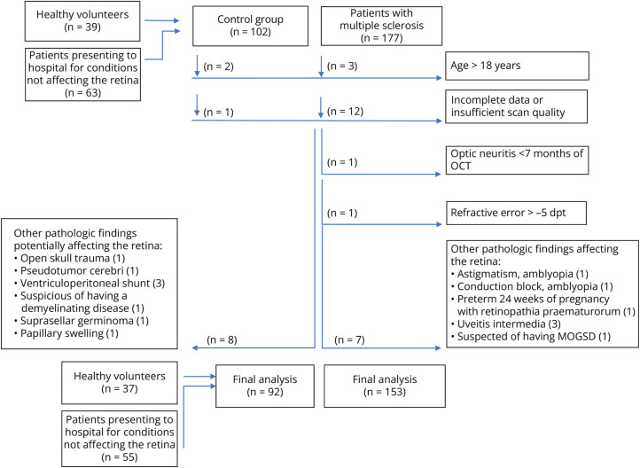
Methods: For this cross-sectional case-control study, we prospectively recruited 153 patients with POMS and 92 controls, including asymptomatic healthy volunteers and children admitted to the hospital with nonretinal disorders. Optical coherence tomography was performed including intraretinal segmentation. Visual function was determined as best corrected visual acuity (BCVA).
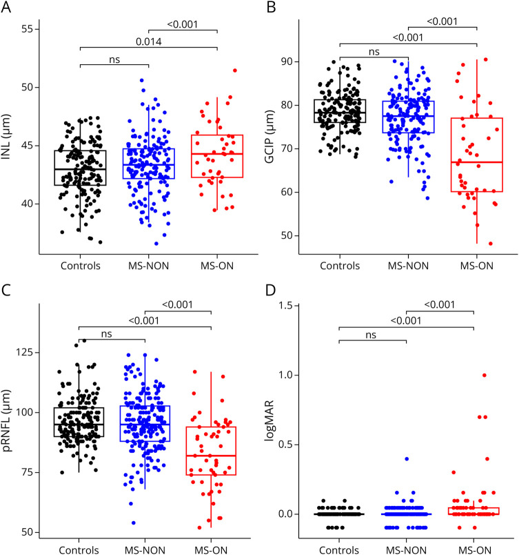
Results: Eyes of children with POMS with prior ON had increased INL thickness (44.31 µm) compared with control eyes (42.96 µm, p = 0.014), whereas pRNFL (83 µm, p < 0.001) and GCIP thickness (68.42 µm, p < 0.001) were reduced compared with control eyes (pRNFL 97 µm, GCIP 78.53 µm). In eyes without history of ON, INL and other layer thicknesses were not different from controls. pRNFL (B = -2, p < 0.001) and GCIP loss (B = -1.6, p < 0.001), but not INL, were associated with worse BCVA. We found macular microcysts in 1 eye of 1 patient with a history of severe ON (0.3%). INL thickness was not associated with age, sex, disease duration, immunotherapy, disability or the MRI parameters T2 lesion count, T2 lesion volume, contrast-enhancing lesions, or contrast-enhancing lesion volume.
Discussion: The INL in POMS shows changes similar to what has been reported in adults, with macular microcysts being much rarer. A lack of cross-sectional association between INL thickness and disease severity may represent the early disease stage with neuroinflammation instead of neurodegeneration being in focus.
背景和目的:小儿发病多发性硬化症(POMS)导致视神经和视网膜损伤,视神经炎(ON)和潜在的亚临床疾病活动。神经轴突性视网膜损伤表现为乳头周围视网膜神经纤维层(pRNFL)和黄斑神经节细胞及内丛状层(GCIP)变薄。内核层(INL)厚度随炎症活动或急性ON后增加,随慢性神经退行性变而减少。成人多发性硬化症患者的视网膜内斑微囊已被发现。本研究的目的是在一个大队列的POMS患者中研究视网膜内斑微囊作为评估病程和治疗成功的潜在生物标志物。方法:在这项横断面病例对照研究中,我们前瞻性地招募了153名POMS患者和92名对照组,包括无症状的健康志愿者和入院的非视网膜疾病儿童。进行光学相干断层扫描,包括视网膜内分割。视功能判定为最佳矫正视力(BCVA)。结果:有ON病史的POMS患儿的眼INL厚度(44.31µm)较对照组(42.96µm, p = 0.014)增加,而pRNFL(83µm, p < 0.001)和GCIP厚度(68.42µm, p < 0.001)较对照组(pRNFL 97µm, GCIP 78.53µm)减少。在无ON病史的眼中,INL及其他层厚度与对照组无差异。pRNFL (B = -2, p < 0.001)和GCIP丢失(B = -1.6, p < 0.001)与BCVA恶化相关,但与INL无关。我们在1例有严重ON病史的患者的1只眼中发现黄斑微囊(0.3%)。INL厚度与年龄、性别、病程、免疫治疗、残疾或MRI参数T2病变计数、T2病变体积、增强病变或增强病变体积无关。讨论:POMS患者的INL表现出与成人相似的变化,黄斑微囊肿更为罕见。INL厚度与疾病严重程度之间缺乏横断面相关性可能代表疾病早期以神经炎症而非神经退行性变为重点。
期刊介绍:
Neurology Neuroimmunology & Neuroinflammation is an official journal of the American Academy of Neurology. Neurology: Neuroimmunology & Neuroinflammation will be the premier peer-reviewed journal in neuroimmunology and neuroinflammation. This journal publishes rigorously peer-reviewed open-access reports of original research and in-depth reviews of topics in neuroimmunology & neuroinflammation, affecting the full range of neurologic diseases including (but not limited to) Alzheimer's disease, Parkinson's disease, ALS, tauopathy, and stroke; multiple sclerosis and NMO; inflammatory peripheral nerve and muscle disease, Guillain-Barré and myasthenia gravis; nervous system infection; paraneoplastic syndromes, noninfectious encephalitides and other antibody-mediated disorders; and psychiatric and neurodevelopmental disorders. Clinical trials, instructive case reports, and small case series will also be featured.

 求助内容:
求助内容: 应助结果提醒方式:


