Sam Springer, Jeremy Basset-Sagarminaga, Tineke van de Weijer, Vera B Schrauwen-Hinderling, Walter H Backes, Roel Wierts
{"title":"Improving image reconstruction to quantify dynamic whole-body PET/CT: Q.Clear versus OSEM.","authors":"Sam Springer, Jeremy Basset-Sagarminaga, Tineke van de Weijer, Vera B Schrauwen-Hinderling, Walter H Backes, Roel Wierts","doi":"10.1186/s40658-025-00736-5","DOIUrl":null,"url":null,"abstract":"<p><strong>Background: </strong>The introduction of PET systems featuring increased count rate sensitivity has resulted in the development of dynamic whole-body PET acquisition protocols to assess <sup>18</sup>F-FDG uptake rate ( <math><msub><mi>K</mi> <mi>i</mi></msub> </math> ) using <sup>18</sup>F-FDG PET/CT. However, in short-axis field-of-view (SAFOV) PET/CT systems, multiple bed positions are required per time frame to achieve whole-body coverage. This results in high noise levels, requiring higher <sup>18</sup>F-FDG activity administration and, consequently, increased patient radiation dose. Bayesian penalized-likelihood PET reconstruction (e.g. Q.Clear, GE Healthcare) has been shown to effectively suppress image noise compared to standard reconstruction techniques. This study investigated the impact of Bayesian penalized-likelihood reconstruction on dynamic whole-body <sup>18</sup>F-FDG PET quantification.</p><p><strong>Methods: </strong>Dynamic whole-body <sup>18</sup>F-FDG PET/CT data (SAFOV PET Discovery MI 5R, GE Healthcare) of healthy volunteers and one lung cancer patient, consisting of a ten-minute dynamic scan of the thoracic region followed by six whole-body passes, were reconstructed with Q.Clear and Ordered Subset Expectation Maximization (OSEM) according to EARL 2 standards. Image noise in the measured time-activity-curves (TAC) was determined for the myocardium, hamstring, liver, subcutaneous adipose tissue and lung lesion for both reconstruction methods. <math><msub><mi>K</mi> <mi>i</mi></msub> </math> values were calculated using Patlak analysis. Finally, bootstrapping was used to investigate the effect of image noise levels on <math><msub><mi>K</mi> <mi>i</mi></msub> </math> values (bias and precision) as a function of magnitude of <math><msub><mi>K</mi> <mi>i</mi></msub> </math> and volume-of-interest (VOI) size for both computationally simulated TACs ( <math><msub><mi>K</mi> <mi>i</mi></msub> </math> = 1.0-50.0·10<sup>-3</sup>·ml·cm<sup>-3</sup>·min<sup>-1</sup>) and the measured TACs.</p><p><strong>Results: </strong>Compared to OSEM, Q.Clear showed 40-55% lower noise levels for all tissue types (p < 0.05). For the measured TACs no systematic bias in <math><msub><mi>K</mi> <mi>i</mi></msub> </math> with either reconstruction method was observed. <math><msub><mi>K</mi> <mi>i</mi></msub> </math> precision decreased with decreasing VOI size, with that of Q.Clear being superior compared to OSEM for small VOIs of 0.56 cm<sup>3</sup> in all tissues (p < 0.05), with the largest difference in relative precision for small values of <math><msub><mi>K</mi> <mi>i</mi></msub> </math> . The simulated TACs corroborated these results, with Q.Clear providing the best precision for small values of <math><msub><mi>K</mi> <mi>i</mi></msub> </math> and small VOIs in all tissues.</p><p><strong>Conclusion: </strong>Q.Clear reconstruction of dynamic whole-body PET/CT data yields more precise <math><msub><mi>K</mi> <mi>i</mi></msub> </math> values, especially for small values of <math><msub><mi>K</mi> <mi>i</mi></msub> </math> and smaller VOIs, compared to standard OSEM. This precision improvement shows Q.Clear's potential to better detect and characterize small lesion metabolic activity in oncology and allows for lower administered activity dosage.</p>","PeriodicalId":11559,"journal":{"name":"EJNMMI Physics","volume":"12 1","pages":"27"},"PeriodicalIF":3.2000,"publicationDate":"2025-03-27","publicationTypes":"Journal Article","fieldsOfStudy":null,"isOpenAccess":false,"openAccessPdf":"https://www.ncbi.nlm.nih.gov/pmc/articles/PMC11947397/pdf/","citationCount":"0","resultStr":null,"platform":"Semanticscholar","paperid":null,"PeriodicalName":"EJNMMI Physics","FirstCategoryId":"3","ListUrlMain":"https://doi.org/10.1186/s40658-025-00736-5","RegionNum":2,"RegionCategory":"医学","ArticlePicture":[],"TitleCN":null,"AbstractTextCN":null,"PMCID":null,"EPubDate":"","PubModel":"","JCR":"Q2","JCRName":"RADIOLOGY, NUCLEAR MEDICINE & MEDICAL IMAGING","Score":null,"Total":0}
引用次数: 0
Abstract
Background: The introduction of PET systems featuring increased count rate sensitivity has resulted in the development of dynamic whole-body PET acquisition protocols to assess 18F-FDG uptake rate ( ) using 18F-FDG PET/CT. However, in short-axis field-of-view (SAFOV) PET/CT systems, multiple bed positions are required per time frame to achieve whole-body coverage. This results in high noise levels, requiring higher 18F-FDG activity administration and, consequently, increased patient radiation dose. Bayesian penalized-likelihood PET reconstruction (e.g. Q.Clear, GE Healthcare) has been shown to effectively suppress image noise compared to standard reconstruction techniques. This study investigated the impact of Bayesian penalized-likelihood reconstruction on dynamic whole-body 18F-FDG PET quantification.
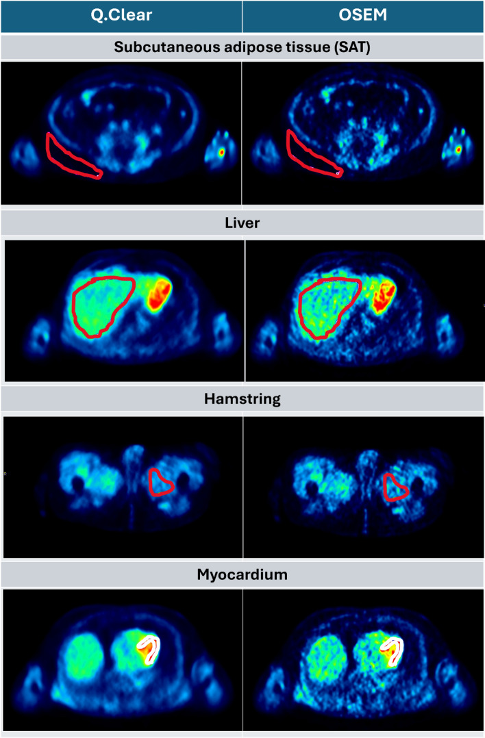
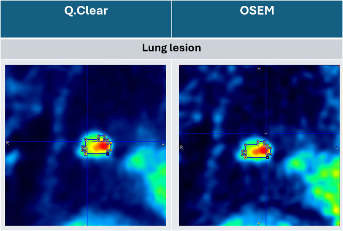
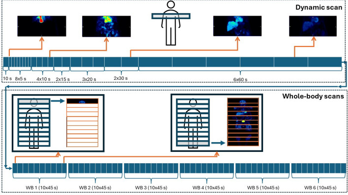
Methods: Dynamic whole-body 18F-FDG PET/CT data (SAFOV PET Discovery MI 5R, GE Healthcare) of healthy volunteers and one lung cancer patient, consisting of a ten-minute dynamic scan of the thoracic region followed by six whole-body passes, were reconstructed with Q.Clear and Ordered Subset Expectation Maximization (OSEM) according to EARL 2 standards. Image noise in the measured time-activity-curves (TAC) was determined for the myocardium, hamstring, liver, subcutaneous adipose tissue and lung lesion for both reconstruction methods. values were calculated using Patlak analysis. Finally, bootstrapping was used to investigate the effect of image noise levels on values (bias and precision) as a function of magnitude of and volume-of-interest (VOI) size for both computationally simulated TACs ( = 1.0-50.0·10-3·ml·cm-3·min-1) and the measured TACs.
Results: Compared to OSEM, Q.Clear showed 40-55% lower noise levels for all tissue types (p < 0.05). For the measured TACs no systematic bias in with either reconstruction method was observed. precision decreased with decreasing VOI size, with that of Q.Clear being superior compared to OSEM for small VOIs of 0.56 cm3 in all tissues (p < 0.05), with the largest difference in relative precision for small values of . The simulated TACs corroborated these results, with Q.Clear providing the best precision for small values of and small VOIs in all tissues.
Conclusion: Q.Clear reconstruction of dynamic whole-body PET/CT data yields more precise values, especially for small values of and smaller VOIs, compared to standard OSEM. This precision improvement shows Q.Clear's potential to better detect and characterize small lesion metabolic activity in oncology and allows for lower administered activity dosage.
背景:PET系统的引入增加了计数率敏感性,导致了动态全身PET采集协议的发展,使用18F-FDG PET/CT评估18F-FDG摄取率(K i)。然而,在短轴视场(SAFOV) PET/CT系统中,每个时间框架需要多个床位才能实现全身覆盖。这导致高噪声水平,需要更高的18F-FDG活性管理,从而增加患者的辐射剂量。与标准重建技术相比,贝叶斯惩罚似然PET重建(如Q.Clear, GE Healthcare)已被证明可以有效地抑制图像噪声。本研究探讨了贝叶斯惩罚似然重建对动态全身18F-FDG PET定量的影响。方法:对健康志愿者和1例肺癌患者的动态全身18F-FDG PET/CT数据(SAFOV PET Discovery MI 5R, GE Healthcare),包括10分钟的胸部区域动态扫描和6次全身扫描,根据EARL 2标准使用Q.Clear and Ordered子集期望最大化(OSEM)进行重建。测定两种重建方法的心肌、腿筋、肝脏、皮下脂肪组织和肺部病变的测量时间-活动曲线(TAC)中的图像噪声。ki值采用Patlak分析法计算。最后,采用自举法研究了图像噪声水平对计算模拟tac (K i = 1.0-50.0·10-3·ml·cm-3·min-1)和测量tac的K i值(偏差和精度)的影响,并将其作为K i大小和感兴趣体积(VOI)大小的函数。结果:与OSEM相比,Q.Clear显示所有组织类型的噪声水平降低40-55% (p K i与任何一种重建方法都观察到)。随着VOI大小的减小,K - i精度降低,在所有组织中,对于0.56 cm3的小VOI, Q.Clear的精度优于OSEM (p K - i)。模拟的tac证实了这些结果,Q.Clear为所有组织的小K i值和小VOIs提供了最好的精度。结论:与标准OSEM相比,动态全身PET/CT数据的清晰重建可获得更精确的K i值,特别是较小的K i值和较小的voi。这种精度的提高显示了Q.Clear在更好地检测和表征肿瘤小病变代谢活动方面的潜力,并允许更低的给药活性剂量。
期刊介绍:
EJNMMI Physics is an international platform for scientists, users and adopters of nuclear medicine with a particular interest in physics matters. As a companion journal to the European Journal of Nuclear Medicine and Molecular Imaging, this journal has a multi-disciplinary approach and welcomes original materials and studies with a focus on applied physics and mathematics as well as imaging systems engineering and prototyping in nuclear medicine. This includes physics-driven approaches or algorithms supported by physics that foster early clinical adoption of nuclear medicine imaging and therapy.

 求助内容:
求助内容: 应助结果提醒方式:


