Magnetic resonance imaging and ultrasound examination in preoperative pelvic staging of early-stage cervical cancer: post-hoc analysis of SENTIX study.

IF 6.3 1区 医学 Q1 ACOUSTICS
Ultrasound in Obstetrics & Gynecology Pub Date : 2025-04-01 Epub Date: 2025-03-25 DOI:10.1002/uog.29205
D Cibula, C Köhler, J Jarkovský, R Kocián, P Dundr, J Klát, I Zapardiel, F Landoni, F Frühauf, R Fischbach, M Borčinová, D Fischerová
{"title":"Magnetic resonance imaging and ultrasound examination in preoperative pelvic staging of early-stage cervical cancer: post-hoc analysis of SENTIX study.","authors":"D Cibula, C Köhler, J Jarkovský, R Kocián, P Dundr, J Klát, I Zapardiel, F Landoni, F Frühauf, R Fischbach, M Borčinová, D Fischerová","doi":"10.1002/uog.29205","DOIUrl":null,"url":null,"abstract":"<p><strong>Objectives: </strong>SENTIX was a prospective, single-arm, international multicenter study that evaluated sentinel lymph node biopsy without pelvic lymph node dissection in patients with early-stage cervical cancer. We aimed to evaluate the concordance between preoperative imaging modalities (magnetic resonance imaging (MRI) and ultrasound) and final pathology in the clinical staging of early-stage cervical cancer by post-hoc analysis of the SENTIX study data.</p><p><strong>Methods: </strong>In total, 47 sites across 18 countries participated in the SENTIX study. Patients with Stage IA1/lymphovascular space invasion-positive to IB2 (International Federation of Gynecology and Obstetrics (FIGO) classification (2018)) cervical cancer, with usual histological types and no suspicious lymph nodes on imaging, were prospectively enrolled between May 2016 and October 2020. Preoperative pelvic clinical staging on either pelvic MRI or ultrasound examination was mandatory. Tumor size discrepancy (< 10 mm vs ≥ 10 mm) between imaging and pathology, as well as the negative predictive value (NPV) of MRI and ultrasound for parametrial involvement and lymph node macrometastasis, were analyzed.</p><p><strong>Results: </strong>Among 690 eligible prospectively enrolled patients, MRI and ultrasound were used as the staging imaging modality in 322 (46.7%) and 298 (43.2%) patients, respectively. A discrepancy of tumor size ≥ 10 mm was reported between ultrasound and final pathology in 39/298 (13.1%) patients and between MRI and pathology in 53/322 (16.5%), with no significant difference in the accuracy of tumor measurement between the two imaging modalities. The NPV of ultrasound in assessing parametrial infiltration and lymph node involvement was 97.0% (95% CI, 0.95-0.99%) and 94.0% (95% CI, 0.91-0.97%), respectively, and that of MRI was 95.3% (95% CI, 0.93-0.98%) and 94.1% (95% CI, 0.92-0.97%), respectively, with no significant differences between the parameters. Ultrasound and MRI were comparable regarding the tumor size measurement (P = 0.452), failure to detect parametrial involvement (P = 0.624) and failure to detect macrometastases in sentinel lymph node (P = 0.876).</p><p><strong>Conclusions: </strong>Pelvic ultrasound examination and MRI had similar concordance with histology in the assessment of tumor size and of parametrial and lymph node invasion in early-stage cervical cancer. Ultrasound examination should be considered part of preoperative pelvic clinical staging in early-stage cervical cancer, especially in limited-resource regions where MRI is unavailable. © 2025 The Author(s). Ultrasound in Obstetrics & Gynecology published by John Wiley & Sons Ltd on behalf of International Society of Ultrasound in Obstetrics and Gynecology.</p>","PeriodicalId":23454,"journal":{"name":"Ultrasound in Obstetrics & Gynecology","volume":" ","pages":"495-502"},"PeriodicalIF":6.3000,"publicationDate":"2025-04-01","publicationTypes":"Journal Article","fieldsOfStudy":null,"isOpenAccess":false,"openAccessPdf":"https://www.ncbi.nlm.nih.gov/pmc/articles/PMC11961100/pdf/","citationCount":"0","resultStr":null,"platform":"Semanticscholar","paperid":null,"PeriodicalName":"Ultrasound in Obstetrics & Gynecology","FirstCategoryId":"3","ListUrlMain":"https://doi.org/10.1002/uog.29205","RegionNum":1,"RegionCategory":"医学","ArticlePicture":[],"TitleCN":null,"AbstractTextCN":null,"PMCID":null,"EPubDate":"2025/3/25 0:00:00","PubModel":"Epub","JCR":"Q1","JCRName":"ACOUSTICS","Score":null,"Total":0}
引用次数: 0

Abstract

Objectives: SENTIX was a prospective, single-arm, international multicenter study that evaluated sentinel lymph node biopsy without pelvic lymph node dissection in patients with early-stage cervical cancer. We aimed to evaluate the concordance between preoperative imaging modalities (magnetic resonance imaging (MRI) and ultrasound) and final pathology in the clinical staging of early-stage cervical cancer by post-hoc analysis of the SENTIX study data.

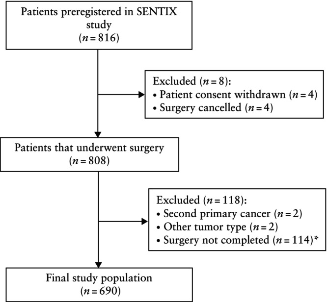
Methods: In total, 47 sites across 18 countries participated in the SENTIX study. Patients with Stage IA1/lymphovascular space invasion-positive to IB2 (International Federation of Gynecology and Obstetrics (FIGO) classification (2018)) cervical cancer, with usual histological types and no suspicious lymph nodes on imaging, were prospectively enrolled between May 2016 and October 2020. Preoperative pelvic clinical staging on either pelvic MRI or ultrasound examination was mandatory. Tumor size discrepancy (< 10 mm vs ≥ 10 mm) between imaging and pathology, as well as the negative predictive value (NPV) of MRI and ultrasound for parametrial involvement and lymph node macrometastasis, were analyzed.

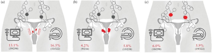
Results: Among 690 eligible prospectively enrolled patients, MRI and ultrasound were used as the staging imaging modality in 322 (46.7%) and 298 (43.2%) patients, respectively. A discrepancy of tumor size ≥ 10 mm was reported between ultrasound and final pathology in 39/298 (13.1%) patients and between MRI and pathology in 53/322 (16.5%), with no significant difference in the accuracy of tumor measurement between the two imaging modalities. The NPV of ultrasound in assessing parametrial infiltration and lymph node involvement was 97.0% (95% CI, 0.95-0.99%) and 94.0% (95% CI, 0.91-0.97%), respectively, and that of MRI was 95.3% (95% CI, 0.93-0.98%) and 94.1% (95% CI, 0.92-0.97%), respectively, with no significant differences between the parameters. Ultrasound and MRI were comparable regarding the tumor size measurement (P = 0.452), failure to detect parametrial involvement (P = 0.624) and failure to detect macrometastases in sentinel lymph node (P = 0.876).

Conclusions: Pelvic ultrasound examination and MRI had similar concordance with histology in the assessment of tumor size and of parametrial and lymph node invasion in early-stage cervical cancer. Ultrasound examination should be considered part of preoperative pelvic clinical staging in early-stage cervical cancer, especially in limited-resource regions where MRI is unavailable. © 2025 The Author(s). Ultrasound in Obstetrics & Gynecology published by John Wiley & Sons Ltd on behalf of International Society of Ultrasound in Obstetrics and Gynecology.

早期宫颈癌术前盆腔分期的磁共振成像和超声检查:SENTIX研究的事后分析。
目的:SENTIX是一项前瞻性、单臂、国际多中心研究,评估早期宫颈癌患者前哨淋巴结活检而不进行盆腔淋巴结清扫。我们旨在通过对SENTIX研究数据的事后分析,评估早期宫颈癌临床分期的术前影像学(磁共振成像(MRI)和超声)与最终病理之间的一致性。方法:共有18个国家的47个站点参与了SENTIX研究。2016年5月至2020年10月,前瞻性纳入IA1期/淋巴血管间隙浸润阳性IB2(国际妇产科联合会(FIGO)分类(2018))宫颈癌患者,组织学类型正常,影像学上无可疑淋巴结。术前盆腔MRI或超声检查的临床分期是强制性的。结果:在690例符合条件的前瞻性入选患者中,分别有322例(46.7%)和298例(43.2%)患者使用MRI和超声作为分期成像方式。39/298例(13.1%)患者超声检查结果与最终病理检查结果存在肿瘤大小≥10 mm的差异,53/322例(16.5%)患者MRI检查结果与最终病理结果存在肿瘤大小≥10 mm的差异,两种成像方式对肿瘤测量的准确性无显著差异。超声评估参数浸润和淋巴结累及的NPV分别为97.0% (95% CI, 0.95-0.99%)和94.0% (95% CI, 0.91-0.97%), MRI评估参数浸润和淋巴结累及的NPV分别为95.3% (95% CI, 0.93-0.98%)和94.1% (95% CI, 0.92-0.97%),参数间无显著差异。超声和MRI在肿瘤大小测量(P = 0.452)、参数累及检测失败(P = 0.624)和前哨淋巴结大转移检测失败(P = 0.876)方面具有可比性。结论:盆腔超声检查和MRI检查对早期宫颈癌的肿瘤大小、参数及淋巴结浸润的评估与组织学具有相似的一致性。超声检查应被视为早期宫颈癌术前盆腔临床分期的一部分,特别是在资源有限且无法获得MRI的地区。©2025作者。妇产科学超声由John Wiley & Sons Ltd代表国际妇产科学超声学会出版。
本文章由计算机程序翻译,如有差异,请以英文原文为准。
求助全文
约1分钟内获得全文 求助全文
来源期刊
CiteScore
12.30
自引率
14.10%
发文量
891
审稿时长
1 months
期刊介绍: Ultrasound in Obstetrics & Gynecology (UOG) is the official journal of the International Society of Ultrasound in Obstetrics and Gynecology (ISUOG) and is considered the foremost international peer-reviewed journal in the field. It publishes cutting-edge research that is highly relevant to clinical practice, which includes guidelines, expert commentaries, consensus statements, original articles, and systematic reviews. UOG is widely recognized and included in prominent abstract and indexing databases such as Index Medicus and Current Contents.
×
引用
GB/T 7714-2015
复制
MLA
复制
APA
复制
导出至
BibTeX EndNote RefMan NoteFirst NoteExpress
×
提示
您的信息不完整,为了账户安全,请先补充。
现在去补充
×
提示
您因"违规操作"
具体请查看互助需知
我知道了
×
提示
确定
请完成安全验证×
copy
已复制链接
快去分享给好友吧!
我知道了
右上角分享
点击右上角分享
0
联系我们:info@booksci.cn Book学术提供免费学术资源搜索服务,方便国内外学者检索中英文文献。致力于提供最便捷和优质的服务体验。 Copyright © 2023 布克学术 All rights reserved.
京ICP备2023020795号-1
ghs 京公网安备 11010802042870号
Book学术文献互助
Book学术文献互助群
群 号:604180095
Book学术官方微信