{"title":"Tumor Budding: A Novel Prognostic Marker in Breast Carcinoma with Correlation of Histopathological and Immunohistochemical Parameters.","authors":"Poornima Manimaran, Ashini Shah, Amisha Gami, Jahnavi Gandhi, Sneha Kakoty, Varnika Rai, Priti P Trivedi","doi":"10.1055/s-0044-1789582","DOIUrl":null,"url":null,"abstract":"<p><strong>Introduction: </strong>Breast cancer is a highly heterogenous tumor with different subtypes showing varying prognosis. Tumor budding is an unfavorable histological feature of many epithelial cancers. The purpose of this study is to analyze the association between tumor bud density with various histological and immunohistochemical characteristics and to explore its prognostic role in breast carcinoma.</p><p><strong>Materials and methods: </strong>A retrospective analysis was performed on 100 patients of breast cancer diagnosed in our institute from January to December 2017. Hematoxylin and eosin (H&E) stained slides from tumors and immunohistochemical slides were reviewed independently by two pathologists, and clinical data were acquired from computerized records. Patients on neoadjuvant chemotherapy were excluded from the study.</p><p><strong>Results: </strong>The study comprised 100 patients of invasive breast carcinoma. The median age was 52 years, and 96% were invasive ductal carcinoma. The median follow-up was 34 months. High tumor bud density was substantially correlated with primary tumor staging (T3, T4; 73% [11/15] cases) and lymph node staging (N2, N3; 68% [13/19] cases) with <i>p</i> -values of 0.017 and 0.023, respectively. Systemic metastasis (85% [6/7] cases) was significantly associated with high tumor bud density ( <i>p</i> =0.025) but lymphovascular invasion (LVI) and perineural invasion (PNI) were not significantly associated with tumor bud density ( <i>p</i> = 0.762 and 0.862, respectively). Patients with N2 nodal stage had low event-free survival rate than N0/N1 nodal stage irrespective of tumor bud status. Grade 3 tumors with high tumor bud density had worse event-free survival than any other grades. There was no association of tumor bud density with tumor staging, necrosis, PNI, LVI, estrogen receptor (ER), progesterone receptor (PR) and <i>Her2/neu</i> , and event-free survival.</p><p><strong>Conclusion: </strong>Strong relationships have been found between tumor bud density and poor prognostic variables such as primary tumor staging and lymph node staging. These results provide credence to the idea that tumor bud density can be an assessable prognostic feature that should be taken into account while reporting breast cancer cases. Tumor bud density evaluation has to be standardized nevertheless if it is to be widely adopted.</p>","PeriodicalId":22053,"journal":{"name":"South Asian Journal of Cancer","volume":"14 1","pages":"38-44"},"PeriodicalIF":0.8000,"publicationDate":"2024-08-28","publicationTypes":"Journal Article","fieldsOfStudy":null,"isOpenAccess":false,"openAccessPdf":"https://www.ncbi.nlm.nih.gov/pmc/articles/PMC11925612/pdf/","citationCount":"0","resultStr":null,"platform":"Semanticscholar","paperid":null,"PeriodicalName":"South Asian Journal of Cancer","FirstCategoryId":"1085","ListUrlMain":"https://doi.org/10.1055/s-0044-1789582","RegionNum":0,"RegionCategory":null,"ArticlePicture":[],"TitleCN":null,"AbstractTextCN":null,"PMCID":null,"EPubDate":"2025/1/1 0:00:00","PubModel":"eCollection","JCR":"Q4","JCRName":"ONCOLOGY","Score":null,"Total":0}
引用次数: 0
Abstract
Introduction: Breast cancer is a highly heterogenous tumor with different subtypes showing varying prognosis. Tumor budding is an unfavorable histological feature of many epithelial cancers. The purpose of this study is to analyze the association between tumor bud density with various histological and immunohistochemical characteristics and to explore its prognostic role in breast carcinoma.
Materials and methods: A retrospective analysis was performed on 100 patients of breast cancer diagnosed in our institute from January to December 2017. Hematoxylin and eosin (H&E) stained slides from tumors and immunohistochemical slides were reviewed independently by two pathologists, and clinical data were acquired from computerized records. Patients on neoadjuvant chemotherapy were excluded from the study.
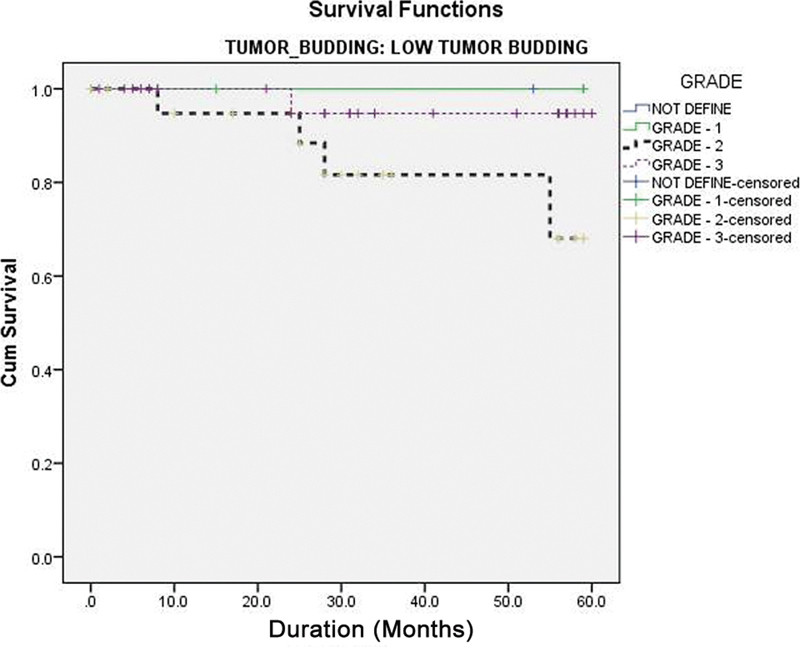
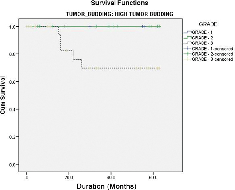
Results: The study comprised 100 patients of invasive breast carcinoma. The median age was 52 years, and 96% were invasive ductal carcinoma. The median follow-up was 34 months. High tumor bud density was substantially correlated with primary tumor staging (T3, T4; 73% [11/15] cases) and lymph node staging (N2, N3; 68% [13/19] cases) with p -values of 0.017 and 0.023, respectively. Systemic metastasis (85% [6/7] cases) was significantly associated with high tumor bud density ( p =0.025) but lymphovascular invasion (LVI) and perineural invasion (PNI) were not significantly associated with tumor bud density ( p = 0.762 and 0.862, respectively). Patients with N2 nodal stage had low event-free survival rate than N0/N1 nodal stage irrespective of tumor bud status. Grade 3 tumors with high tumor bud density had worse event-free survival than any other grades. There was no association of tumor bud density with tumor staging, necrosis, PNI, LVI, estrogen receptor (ER), progesterone receptor (PR) and Her2/neu , and event-free survival.
Conclusion: Strong relationships have been found between tumor bud density and poor prognostic variables such as primary tumor staging and lymph node staging. These results provide credence to the idea that tumor bud density can be an assessable prognostic feature that should be taken into account while reporting breast cancer cases. Tumor bud density evaluation has to be standardized nevertheless if it is to be widely adopted.

 求助内容:
求助内容: 应助结果提醒方式:


