Chinmay Mahatme, Madhurima Kaushik, Veerappan Rathinasabapathy Saravanan, Karthik Kumar, Virna M Shah
{"title":"Macular microvascular and structural changes on optical coherence tomography angiography in atypical optic neuritis.","authors":"Chinmay Mahatme, Madhurima Kaushik, Veerappan Rathinasabapathy Saravanan, Karthik Kumar, Virna M Shah","doi":"10.5662/wjm.v15.i1.98482","DOIUrl":null,"url":null,"abstract":"<p><strong>Background: </strong>Atypical optic neuritis, consisting of neuromyelitis optica spectrum disorders (NMOSD) or myelin oligodendrocyte glycoprotein antibody disease (MOGAD), has a very similar presentation but different prognostic implications and long-term management strategies. Vascular and metabolic factors are being thought to play a role in such autoimmune neuro-inflammatory disorders, apart from the obvious immune mediated damage. With the advent of optical coherence tomography angiography (OCTA), it is easy to pick up on these subclinical macular microvascular and structural changes.</p><p><strong>Aim: </strong>To study the macular microvascular and structural changes on OCTA in atypical optic neuritis.</p><p><strong>Methods: </strong>This observational cross-sectional study involved 8 NMOSD and 17 MOGAD patients, diagnosed serologically, as well as 10 healthy controls. Macular vascular density (MVD) and ganglion cell + inner plexiform layer thickness (GCIPL) were studied using OCTA.</p><p><strong>Results: </strong>There was a significant reduction in MVD in NMOSD and MOGAD affected as well as unaffected eyes when compared with healthy controls. NMOSD and MOGAD affected eyes had significant GCIPL thinning compared with healthy controls. NMOSD unaffected eyes did not show significant GCIPL thinning compared to healthy controls in contrast to MOGAD unaffected eyes. On comparing NMOSD with MOGAD, there was no significant difference in terms of MVD or GCIPL in the affected or unaffected eyes.</p><p><strong>Conclusion: </strong>Although significant microvascular and structural changes are present on OCTA between atypical optic neuritis and normal patients, they could not help in differentiating between NMOSD and MOGAD cases.</p>","PeriodicalId":94271,"journal":{"name":"World journal of methodology","volume":"15 1","pages":"98482"},"PeriodicalIF":0.0000,"publicationDate":"2025-03-20","publicationTypes":"Journal Article","fieldsOfStudy":null,"isOpenAccess":false,"openAccessPdf":"https://www.ncbi.nlm.nih.gov/pmc/articles/PMC11525885/pdf/","citationCount":"0","resultStr":null,"platform":"Semanticscholar","paperid":null,"PeriodicalName":"World journal of methodology","FirstCategoryId":"1085","ListUrlMain":"https://doi.org/10.5662/wjm.v15.i1.98482","RegionNum":0,"RegionCategory":null,"ArticlePicture":[],"TitleCN":null,"AbstractTextCN":null,"PMCID":null,"EPubDate":"","PubModel":"","JCR":"","JCRName":"","Score":null,"Total":0}
引用次数: 0
Abstract
Background: Atypical optic neuritis, consisting of neuromyelitis optica spectrum disorders (NMOSD) or myelin oligodendrocyte glycoprotein antibody disease (MOGAD), has a very similar presentation but different prognostic implications and long-term management strategies. Vascular and metabolic factors are being thought to play a role in such autoimmune neuro-inflammatory disorders, apart from the obvious immune mediated damage. With the advent of optical coherence tomography angiography (OCTA), it is easy to pick up on these subclinical macular microvascular and structural changes.
Aim: To study the macular microvascular and structural changes on OCTA in atypical optic neuritis.
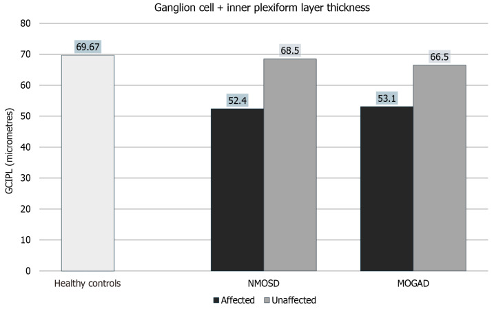
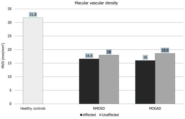
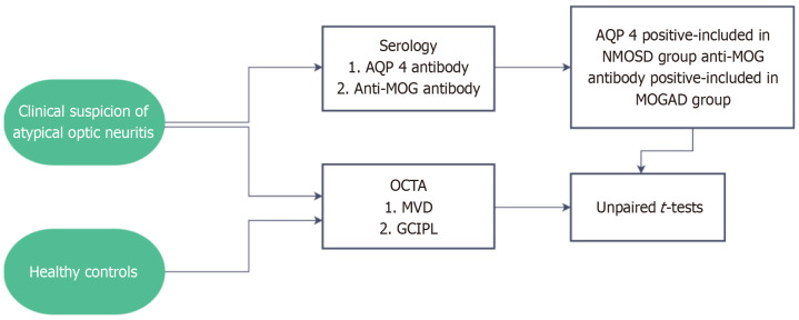
Methods: This observational cross-sectional study involved 8 NMOSD and 17 MOGAD patients, diagnosed serologically, as well as 10 healthy controls. Macular vascular density (MVD) and ganglion cell + inner plexiform layer thickness (GCIPL) were studied using OCTA.
Results: There was a significant reduction in MVD in NMOSD and MOGAD affected as well as unaffected eyes when compared with healthy controls. NMOSD and MOGAD affected eyes had significant GCIPL thinning compared with healthy controls. NMOSD unaffected eyes did not show significant GCIPL thinning compared to healthy controls in contrast to MOGAD unaffected eyes. On comparing NMOSD with MOGAD, there was no significant difference in terms of MVD or GCIPL in the affected or unaffected eyes.
Conclusion: Although significant microvascular and structural changes are present on OCTA between atypical optic neuritis and normal patients, they could not help in differentiating between NMOSD and MOGAD cases.

 求助内容:
求助内容: 应助结果提醒方式:


