Eduardo De Faria Castro Fleury, Michele Rodrigues da Silva Junqueira, Tiago Sarmet Esteves Teixeira, Pryscilla Alves Ferreira, Giulia Matheus E Castro, Bruna Aguiar Portugal Viotti
{"title":"Dedicated Ultrasonography Versus Magnetic Resonance Imaging for Breast Implant Evaluation, Initial Study.","authors":"Eduardo De Faria Castro Fleury, Michele Rodrigues da Silva Junqueira, Tiago Sarmet Esteves Teixeira, Pryscilla Alves Ferreira, Giulia Matheus E Castro, Bruna Aguiar Portugal Viotti","doi":"10.2147/MDER.S503466","DOIUrl":null,"url":null,"abstract":"<p><strong>Aim: </strong>Describe the role of dedicated ultrasound in evaluating breast implants compared to breast magnetic resonance.</p><p><strong>Materials and methods: </strong>A prospective observational study was conducted in a single center to evaluate breast implant complications in patients referred to breast magnetic resonance (MRI) scan. All patients who had breast implants submitted to MRI scan were invited to a complementary dedicated ultrasound (US) evaluation of the breast implants. The implant changes were classified following a dedicated protocol. The classifiers used to evaluate the implant include evaluation of implant surface (shell), implant internal content homogeneity, fibrous capsule, intracapsular space, pericapsular space, and axillary extension.</p><p><strong>Results: </strong>Preliminary results included evaluating 29 consecutive patients who underwent MRI. Twenty-nine patients with 49 implants were included in the study. The US showed a superior ability to assess the internal contents of the implants, the implant surface, and the intracapsular contents. There were no significant statistical differences in evaluating macro changes such as implant location, intracapsular collection, and radio-frequency identification (RFID) presence. MRI was superior to the US in classifying the fibrous capsule type.</p><p><strong>Conclusion: </strong>US of silicone implants can be used as an alternative to MRI to evaluate implant complications.</p>","PeriodicalId":47140,"journal":{"name":"Medical Devices-Evidence and Research","volume":"18 ","pages":"177-189"},"PeriodicalIF":1.5000,"publicationDate":"2025-03-14","publicationTypes":"Journal Article","fieldsOfStudy":null,"isOpenAccess":false,"openAccessPdf":"https://www.ncbi.nlm.nih.gov/pmc/articles/PMC11920629/pdf/","citationCount":"0","resultStr":null,"platform":"Semanticscholar","paperid":null,"PeriodicalName":"Medical Devices-Evidence and Research","FirstCategoryId":"1085","ListUrlMain":"https://doi.org/10.2147/MDER.S503466","RegionNum":0,"RegionCategory":null,"ArticlePicture":[],"TitleCN":null,"AbstractTextCN":null,"PMCID":null,"EPubDate":"2025/1/1 0:00:00","PubModel":"eCollection","JCR":"Q4","JCRName":"ENGINEERING, BIOMEDICAL","Score":null,"Total":0}
引用次数: 0
Abstract
Aim: Describe the role of dedicated ultrasound in evaluating breast implants compared to breast magnetic resonance.
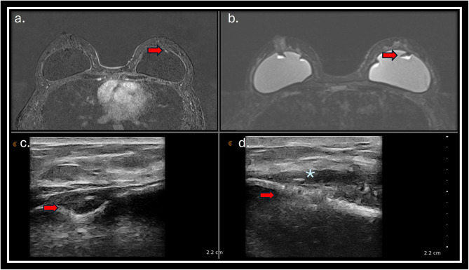
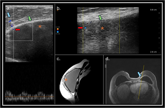
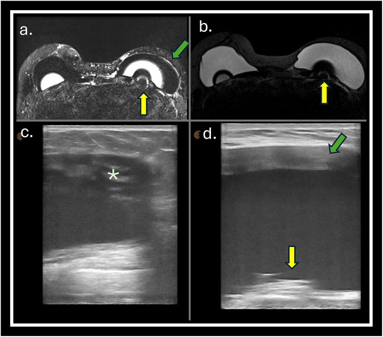
Materials and methods: A prospective observational study was conducted in a single center to evaluate breast implant complications in patients referred to breast magnetic resonance (MRI) scan. All patients who had breast implants submitted to MRI scan were invited to a complementary dedicated ultrasound (US) evaluation of the breast implants. The implant changes were classified following a dedicated protocol. The classifiers used to evaluate the implant include evaluation of implant surface (shell), implant internal content homogeneity, fibrous capsule, intracapsular space, pericapsular space, and axillary extension.
Results: Preliminary results included evaluating 29 consecutive patients who underwent MRI. Twenty-nine patients with 49 implants were included in the study. The US showed a superior ability to assess the internal contents of the implants, the implant surface, and the intracapsular contents. There were no significant statistical differences in evaluating macro changes such as implant location, intracapsular collection, and radio-frequency identification (RFID) presence. MRI was superior to the US in classifying the fibrous capsule type.
Conclusion: US of silicone implants can be used as an alternative to MRI to evaluate implant complications.

 求助内容:
求助内容: 应助结果提醒方式:


