Lovisa E L Westlund Gotby, Martina Stella, Camille D E Van Speybroeck, Daphne Lobeek, Floris H P van Velden, Mette K Stam, Petra Dibbets-Schneider, Daphne M V de Vries-Huizing, Erik-Jan Rijkhorst, Berlinda J de Wit-van de Veen, Roel Wierts, Rob van Rooij
{"title":"Towards harmonized holmium-166 SPECT image quality for dosimetry: a multi-center, multi-vendor study.","authors":"Lovisa E L Westlund Gotby, Martina Stella, Camille D E Van Speybroeck, Daphne Lobeek, Floris H P van Velden, Mette K Stam, Petra Dibbets-Schneider, Daphne M V de Vries-Huizing, Erik-Jan Rijkhorst, Berlinda J de Wit-van de Veen, Roel Wierts, Rob van Rooij","doi":"10.1186/s40658-025-00733-8","DOIUrl":null,"url":null,"abstract":"<p><strong>Background: </strong>Reliable dosimetry based on SPECT/CT imaging is essential to achieve personalized <sup>166</sup>Ho-radioembolization treatment planning and evaluation. This study quantitatively evaluates multiple acquisition and reconstruction protocols for <sup>166</sup>Ho-SPECT imaging based on data from five Dutch hospitals. We aim to recommend an imaging protocol which harmonizes <sup>166</sup>Ho-SPECT images for reproducible and accurate dosimetry in a multi-scanner and multi-center setting.</p><p><strong>Methods: </strong>Cylindrical and NEMA IEC phantoms, filled with <sup>166</sup>Ho-chloride, were imaged using seven SPECT/CT scanners from two vendors (GE HealthCare and Siemens Healthineers). Data were acquired with a photopeak window centered at 81 keV. Two adjacent scatter windows, and one upper scatter window at 118 keV were used for triple-energy window (TEW) and dual-energy window (DEW) scatter correction, respectively. The TEW and DEW reconstructions used vendor-specific software. Additionally, a vendor-neutral software package with Monte Carlo (MC) scatter correction (Hermes Medical Solutions) was used to study the influence of scanner hardware on the image quality. System sensitivity was measured in projection data of the cylindrical phantom. The axial uniformity in the cylindrical phantom was used to characterize the impact of the scatter correction method. The image quality was evaluated by the coefficient of variation (COV; noise), the contrast recovery coefficients (CRCs) and contrast-to-noise ratios (CNRs).</p><p><strong>Results: </strong>TEW scatter correction resulted in superior uniformity and higher CRCs compared to the DEW (CRC for the largest sphere over all scanners, mean ± SD (range): TEW 0.54 ± 0.07 (0.36-0.65), DEW 0.44 ± 0.04 (0.34-0.51)). DEW resulted in lower noise levels compared to TEW (16% lower on average). The DEW and TEW images resulted in comparable CNRs. The system sensitivities and the vendor-neutral image reconstructions demonstrated differences in hardware between the two vendors, most likely due to the characteristics of the vendor-specific medium energy collimator.</p><p><strong>Conclusion: </strong>This study demonstrates that TEW scatter correction increases the accuracy of <sup>166</sup>Ho-SPECT images compared to DEW, and we henceforth recommend adopting this method in the clinical <sup>166</sup>Ho-dosimetry workflow. Scanner hardware has a substantial impact on the characteristics of the acquired data, and identical reconstruction settings will therefore not automatically lead to harmonized image quality.</p>","PeriodicalId":11559,"journal":{"name":"EJNMMI Physics","volume":"12 1","pages":"24"},"PeriodicalIF":3.2000,"publicationDate":"2025-03-19","publicationTypes":"Journal Article","fieldsOfStudy":null,"isOpenAccess":false,"openAccessPdf":"https://www.ncbi.nlm.nih.gov/pmc/articles/PMC11920561/pdf/","citationCount":"0","resultStr":null,"platform":"Semanticscholar","paperid":null,"PeriodicalName":"EJNMMI Physics","FirstCategoryId":"3","ListUrlMain":"https://doi.org/10.1186/s40658-025-00733-8","RegionNum":2,"RegionCategory":"医学","ArticlePicture":[],"TitleCN":null,"AbstractTextCN":null,"PMCID":null,"EPubDate":"","PubModel":"","JCR":"Q2","JCRName":"RADIOLOGY, NUCLEAR MEDICINE & MEDICAL IMAGING","Score":null,"Total":0}
引用次数: 0
Abstract
Background: Reliable dosimetry based on SPECT/CT imaging is essential to achieve personalized 166Ho-radioembolization treatment planning and evaluation. This study quantitatively evaluates multiple acquisition and reconstruction protocols for 166Ho-SPECT imaging based on data from five Dutch hospitals. We aim to recommend an imaging protocol which harmonizes 166Ho-SPECT images for reproducible and accurate dosimetry in a multi-scanner and multi-center setting.
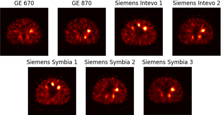
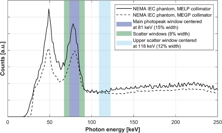
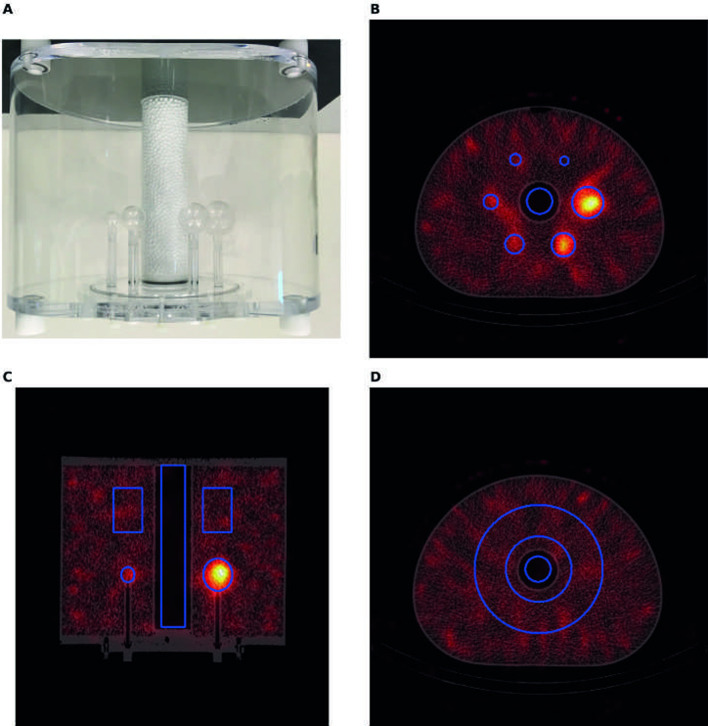
Methods: Cylindrical and NEMA IEC phantoms, filled with 166Ho-chloride, were imaged using seven SPECT/CT scanners from two vendors (GE HealthCare and Siemens Healthineers). Data were acquired with a photopeak window centered at 81 keV. Two adjacent scatter windows, and one upper scatter window at 118 keV were used for triple-energy window (TEW) and dual-energy window (DEW) scatter correction, respectively. The TEW and DEW reconstructions used vendor-specific software. Additionally, a vendor-neutral software package with Monte Carlo (MC) scatter correction (Hermes Medical Solutions) was used to study the influence of scanner hardware on the image quality. System sensitivity was measured in projection data of the cylindrical phantom. The axial uniformity in the cylindrical phantom was used to characterize the impact of the scatter correction method. The image quality was evaluated by the coefficient of variation (COV; noise), the contrast recovery coefficients (CRCs) and contrast-to-noise ratios (CNRs).
Results: TEW scatter correction resulted in superior uniformity and higher CRCs compared to the DEW (CRC for the largest sphere over all scanners, mean ± SD (range): TEW 0.54 ± 0.07 (0.36-0.65), DEW 0.44 ± 0.04 (0.34-0.51)). DEW resulted in lower noise levels compared to TEW (16% lower on average). The DEW and TEW images resulted in comparable CNRs. The system sensitivities and the vendor-neutral image reconstructions demonstrated differences in hardware between the two vendors, most likely due to the characteristics of the vendor-specific medium energy collimator.
Conclusion: This study demonstrates that TEW scatter correction increases the accuracy of 166Ho-SPECT images compared to DEW, and we henceforth recommend adopting this method in the clinical 166Ho-dosimetry workflow. Scanner hardware has a substantial impact on the characteristics of the acquired data, and identical reconstruction settings will therefore not automatically lead to harmonized image quality.
期刊介绍:
EJNMMI Physics is an international platform for scientists, users and adopters of nuclear medicine with a particular interest in physics matters. As a companion journal to the European Journal of Nuclear Medicine and Molecular Imaging, this journal has a multi-disciplinary approach and welcomes original materials and studies with a focus on applied physics and mathematics as well as imaging systems engineering and prototyping in nuclear medicine. This includes physics-driven approaches or algorithms supported by physics that foster early clinical adoption of nuclear medicine imaging and therapy.

 求助内容:
求助内容: 应助结果提醒方式:


