{"title":"Artificial intelligence for predicting interstitial fibrosis and tubular atrophy using diagnostic ultrasound imaging and biomarkers.","authors":"Ting-Wei Chang, Chang-Yu Tsai, Zhen-Yi Tang, Cai-Mei Zheng, Chia-Te Liao, Chung-Yi Cheng, Mai-Szu Wu, Che-Chou Shen, Yen-Chung Lin","doi":"10.1136/bmjhci-2024-101192","DOIUrl":null,"url":null,"abstract":"<p><strong>Background: </strong>Chronic kidney disease (CKD) is a global health concern characterised by irreversible renal damage that is often assessed using invasive renal biopsy. Accurate evaluation of interstitial fibrosis and tubular atrophy (IFTA) is crucial for CKD management. This study aimed to leverage machine learning (ML) models to predict IFTA using a combination of ultrasonography (US) images and patient biomarkers.</p><p><strong>Methods: </strong>We retrospectively collected US images and biomarkers from 632 patients with CKD across three hospitals. The data were subjected to pre-processing, exclusion of sub-optimal images, and feature extraction using a dual-path convolutional neural network. Various ML models, including XGBoost, random forest and logistic regression, were trained and validated using fivefold cross-validation.</p><p><strong>Results: </strong>The dataset was divided into training and test datasets. For image-level IFTA classification, the best performance was achieved by combining US image features and patient biomarkers, with logistic regression yielding an area under the receiver operating characteristic curve (AUROC) of 99%. At the patient level, logistic regression combining US image features and biomarkers provided an AUROC of 96%. Models trained solely on US image features or biomarkers also exhibited high performance, with AUROC exceeding 80%.</p><p><strong>Conclusion: </strong>Our artificial intelligence-based approach to IFTA classification demonstrated high accuracy and AUROC across various ML models. By leveraging patient biomarkers alone, this method offers a non-invasive and robust tool for early CKD assessment, demonstrating that biomarkers alone may suffice for accurate predictions without the added complexity of image-derived features.</p>","PeriodicalId":9050,"journal":{"name":"BMJ Health & Care Informatics","volume":"32 1","pages":""},"PeriodicalIF":4.4000,"publicationDate":"2025-03-17","publicationTypes":"Journal Article","fieldsOfStudy":null,"isOpenAccess":false,"openAccessPdf":"https://www.ncbi.nlm.nih.gov/pmc/articles/PMC11931887/pdf/","citationCount":"0","resultStr":null,"platform":"Semanticscholar","paperid":null,"PeriodicalName":"BMJ Health & Care Informatics","FirstCategoryId":"1085","ListUrlMain":"https://doi.org/10.1136/bmjhci-2024-101192","RegionNum":0,"RegionCategory":null,"ArticlePicture":[],"TitleCN":null,"AbstractTextCN":null,"PMCID":null,"EPubDate":"","PubModel":"","JCR":"Q1","JCRName":"HEALTH CARE SCIENCES & SERVICES","Score":null,"Total":0}
引用次数: 0
Abstract
Background: Chronic kidney disease (CKD) is a global health concern characterised by irreversible renal damage that is often assessed using invasive renal biopsy. Accurate evaluation of interstitial fibrosis and tubular atrophy (IFTA) is crucial for CKD management. This study aimed to leverage machine learning (ML) models to predict IFTA using a combination of ultrasonography (US) images and patient biomarkers.
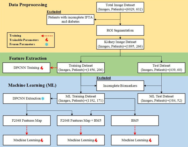
Methods: We retrospectively collected US images and biomarkers from 632 patients with CKD across three hospitals. The data were subjected to pre-processing, exclusion of sub-optimal images, and feature extraction using a dual-path convolutional neural network. Various ML models, including XGBoost, random forest and logistic regression, were trained and validated using fivefold cross-validation.
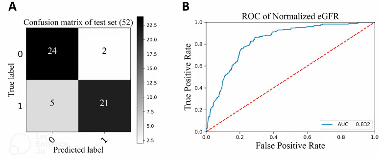
Results: The dataset was divided into training and test datasets. For image-level IFTA classification, the best performance was achieved by combining US image features and patient biomarkers, with logistic regression yielding an area under the receiver operating characteristic curve (AUROC) of 99%. At the patient level, logistic regression combining US image features and biomarkers provided an AUROC of 96%. Models trained solely on US image features or biomarkers also exhibited high performance, with AUROC exceeding 80%.
Conclusion: Our artificial intelligence-based approach to IFTA classification demonstrated high accuracy and AUROC across various ML models. By leveraging patient biomarkers alone, this method offers a non-invasive and robust tool for early CKD assessment, demonstrating that biomarkers alone may suffice for accurate predictions without the added complexity of image-derived features.

 求助内容:
求助内容: 应助结果提醒方式:


